

9.4 Chromosomes and chromosomal aberrations in man, methodology. (a) Numerical and structural aberrations (disorders). (b) Sex chromosomal aberrations – Klinefelter (XXY), Turner (XO), Super female (XXX), intersex and other syndromic disorders. (c) Autosomal aberrations – Down syndrome, Patau, Edward and Cri-du-chat syndromes. (d) Genetic imprints in human disease, genetic screening, genetic counseling, human DNA profiling, gene mapping and genome study.
Official Syllabus: Paper I, Unit 9.4
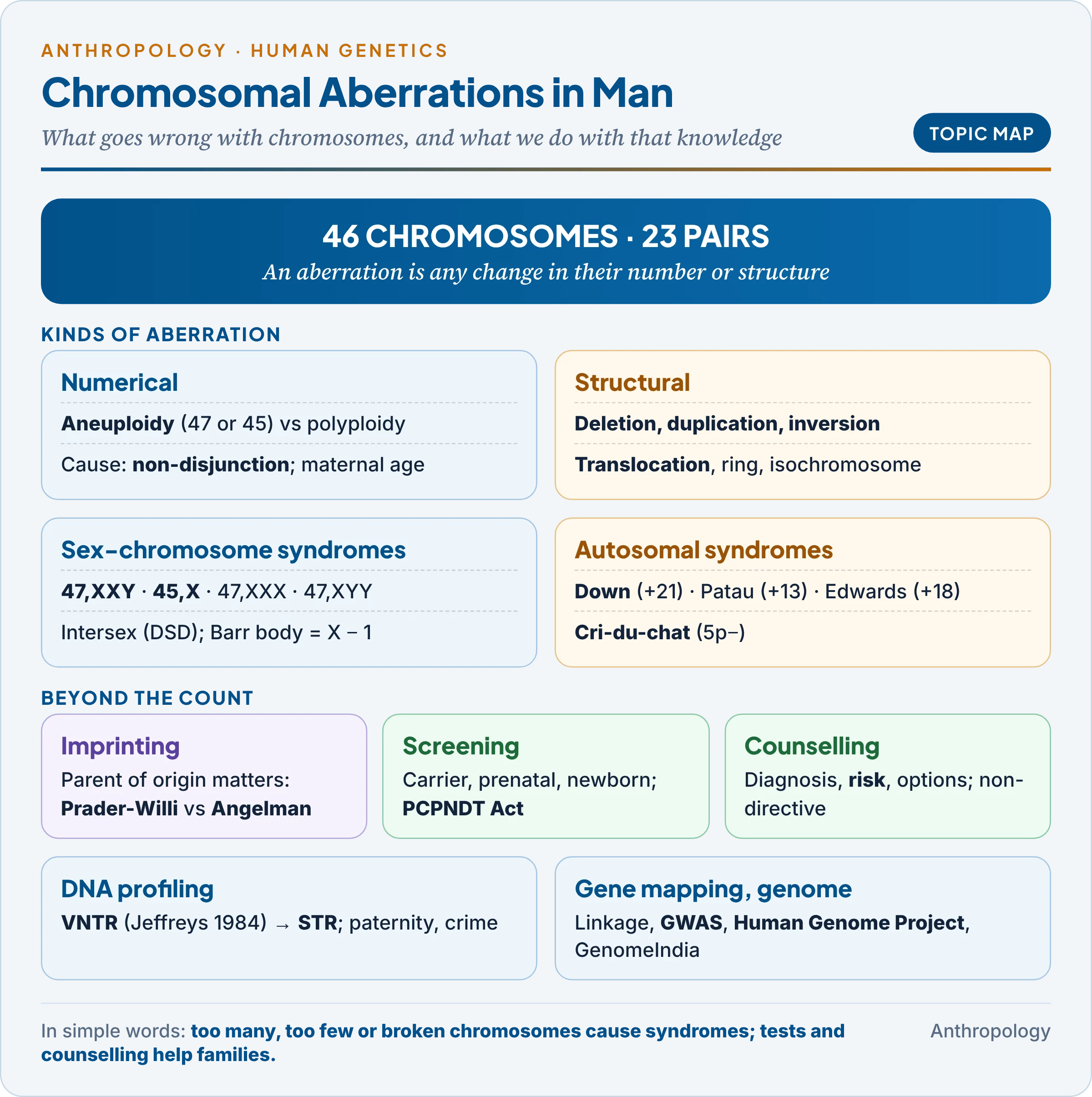
Unit 1.7 described what a chromosome is, and Unit 9.1 how a karyotype is made: blood cells are grown, stopped at metaphase with colchicine, G-banded and arranged in 23 pairs, and written as 46,XY. This unit asks what happens when that picture is wrong (a chromosome too many, one too few, or a broken piece), then follows the uses of this knowledge: screening, counselling, DNA profiling and genome mapping.
1. Chromosomes and the Language of Aberrations
In simple words
Every body cell normally carries 46 chromosomes in 23 pairs. A chromosomal aberration is any change in their number or structure. Because each chromosome carries hundreds of genes, even one extra or missing chromosome upsets the body’s chemistry and usually causes a recognisable syndrome or a miscarriage.
A baby in a district hospital has low muscle tone, a flat face and a single crease across each palm. The karyotype reads 47,XY,+21: a boy with 47 chromosomes, the extra one a chromosome 21, the cause of Down syndrome. Reading such reports needs four ideas.
- Autosomes and sex chromosomes: 22 pairs of autosomes (chromosomes not involved in sex determination) plus XX or XY. The SRY gene (sex-determining region of Y) on Yp turns the embryonic gonad into a testis; without it an ovary forms. So a person with a Y is usually male whatever the number of X chromosomes.
- Centromere position: metacentric (central), submetacentric (off-centre) or acrocentric (near one end: 13, 14, 15, 21, 22); sorted by size into groups A to G (Denver system, 1960).
- Arms and bands: the short arm is p and the long arm q. 1q23 means chromosome 1, long arm, region 2, band 3; bands are numbered outward from the centromere.
- ISCN notation (International System for Human Cytogenomic Nomenclature): number of chromosomes, sex chromosomes, then the change: 47,XX,+21 (trisomy 21), 45,X (Turner), 46,XY,del(5p) or 46,XY,5p− (cri-du-chat), 45,XX,t(14;21) (balanced translocation carrier), 46,XX/47,XX,+21 (a mosaic: two cell lines in one person).
1.1 X-inactivation and the Barr body
The British-Canadian anatomists Murray Barr and Ewart Bertram (1949) saw a dark spot, the sex chromatin or Barr body, in the nuclei of female cells only. The English geneticist Mary Lyon (1961) explained it: in every female cell one X is switched off early in development, at random, and condensed into the Barr body (the Lyon hypothesis).
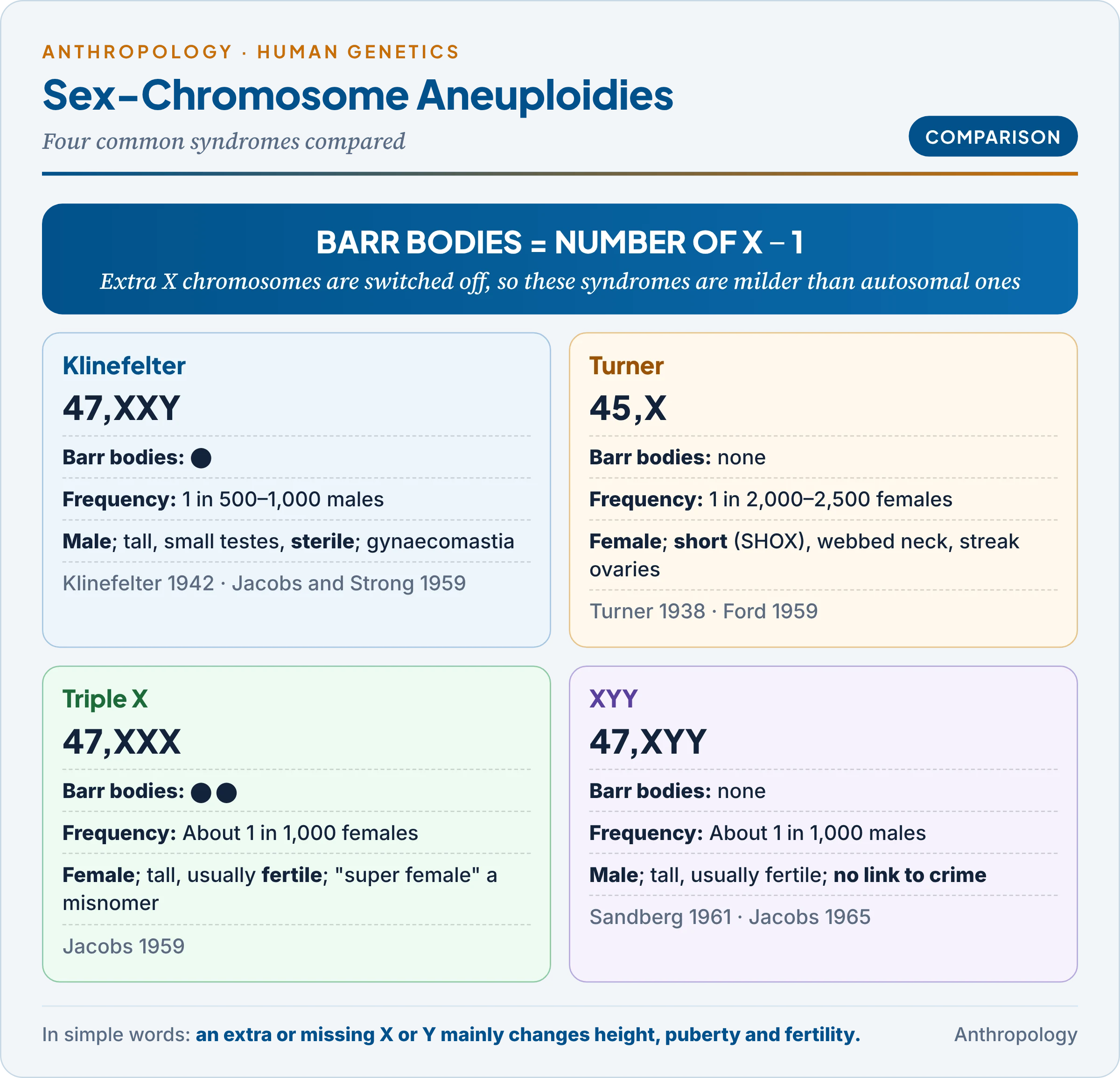
- Rule: number of Barr bodies = number of X chromosomes − 1. Turner (45,X) has none; Klinefelter (47,XXY) has one; 47,XXX has two.
- Why it matters: because extra X chromosomes are silenced, sex-chromosome aneuploidy is far milder than autosomal aneuploidy; symptoms come from the few genes that escape inactivation (such as SHOX, for height).
1.2 Methodology in brief
The laboratory methods were explained in Unit 9.1. The table recalls which method suits which kind of aberration.
| Method | What it sees | Best for |
|---|---|---|
| Karyotype with G-banding (Giemsa) | Whole chromosomes, about 400–550 bands | Aneuploidy, large deletions and translocations (above about 5–10 million bases) |
| Q, R, C, NOR, T banding | Quinacrine fluorescence and other stains | Y (Q), chromosome ends (R, T), heterochromatin (C) |
| FISH (fluorescence in situ hybridisation) | One DNA sequence lit by a fluorescent probe | Microdeletions (22q11.2, 15q11–q13); rapid counting of 13, 18, 21, X, Y in uncultured cells |
| Chromosomal microarray (array CGH) | Gains and losses across the genome | Small copy-number changes; first test for unexplained developmental delay |
| NIPT (non-invasive prenatal testing) | Fetal DNA fragments in the mother’s blood | Screening for trisomies 21, 18, 13 |
Each newer method sees smaller changes, but only the karyotype shows how material is arranged: a balanced translocation, with nothing gained or lost, is invisible to a microarray.
2. Numerical Aberrations
In simple words
A numerical aberration means the cell has the wrong number of chromosomes. It usually arises when a pair of chromosomes fails to separate while eggs or sperm are being made, so that one gamete gets an extra copy and another gets none. The resulting child has 47 or 45 chromosomes instead of 46.
2.1 Types
The terms describe whether whole sets or single chromosomes are affected. Heteroploidy is the general word for any change in number.
| Term | Meaning | Human example |
|---|---|---|
| Euploidy | Exact multiples of the set (n = 23) | Diploid 2n = 46 is normal |
| Polyploidy | Extra whole sets | Triploidy (69,XXY) and tetraploidy (92): found in miscarriages and partial molar pregnancies; not compatible with long survival |
| Aneuploidy | One or a few chromosomes extra or missing | Trisomy (2n+1: 47,+21), monosomy (2n−1: 45,X), tetrasomy (2n+2: 48,XXXY); nullisomy (2n−2) is lethal |
Chromosome errors are common: about half of first-trimester miscarriages carry one, so natural selection removes most before birth.
2.2 Non-disjunction: the main mechanism
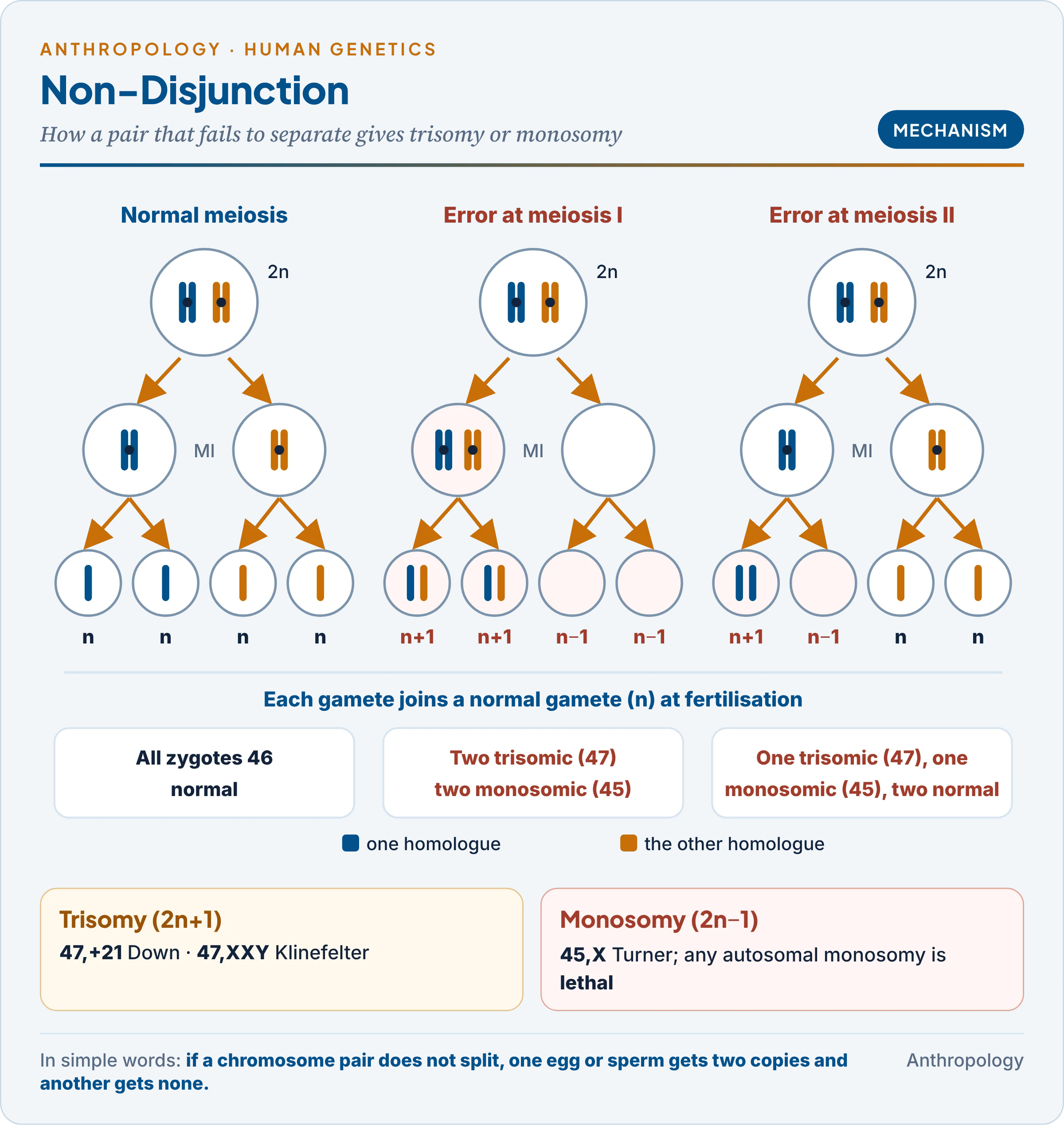
Non-disjunction is the failure of a chromosome pair (meiosis I) or of sister chromatids (meiosis II) to separate at anaphase.
- In meiosis I: both homologues go to one pole, giving two n+1 and two n−1 gametes.
- In meiosis II: one secondary cell is affected, giving one n+1, one n−1 and two normal gametes.
- Fertilisation of an n+1 gamete by a normal one gives a trisomic zygote (47); of an n−1 gamete, a monosomic zygote (45).
- Mitotic non-disjunction after fertilisation gives mosaicism, usually with milder features because only some cells are abnormal.
- Anaphase lag (a chromosome left behind on the spindle and lost) is a second cause of monosomy, common in Turner syndrome.
- Primary and secondary non-disjunction: the first arises in normal parents; the second occurs when a parent already trisomic (or carrying a translocation) passes on the error.
2.3 The maternal age effect
A woman’s oocytes (egg cells) begin meiosis before her own birth and pause in prophase I for 15 to 45 years until ovulation. Over these decades the cohesin proteins holding paired chromosomes weaken, so pairs separate wrongly more often; sperm, by contrast, are made continuously. Hence about 90 per cent of trisomy 21 arises in maternal meiosis, mostly meiosis I, and risk rises steeply with age (§4.1).
Why only a few aneuploidies survive to birth: monosomy of any autosome is lethal in the embryo. Only trisomies of 13, 18 and 21, the chromosomes carrying the fewest genes, reach term, along with the sex-chromosome aneuploidies, which X-inactivation softens. Severity follows gene content: trisomy 21 (fewest genes) is compatible with a long life, trisomy 13 and 18 rarely with one year.
Common confusion
Is aneuploidy the same as polyploidy? No. Polyploidy adds whole sets of 23 (69 or 92 chromosomes). Aneuploidy adds or removes a single chromosome (45 or 47). Down, Turner and Klinefelter syndromes are all aneuploidies; triploidy is polyploidy and ends in miscarriage.
3. Sex-Chromosome Aberrations
In simple words
These conditions involve an extra or missing X or Y chromosome. Because extra X chromosomes are switched off and the Y carries few genes, the effects are milder than those of autosomal errors: people mainly show differences in height, puberty and fertility, and many are never diagnosed.
The four commonest sex-chromosome aneuploidies are compared below; the text after the table explains each.
| Feature | Klinefelter | Turner | Triple X | XYY |
|---|---|---|---|---|
| Karyotype | 47,XXY | 45,X (XO) | 47,XXX | 47,XYY |
| Described | Klinefelter 1942; karyotype Jacobs and Strong 1959 | Turner 1938; karyotype Ford 1959 | Jacobs 1959 | Sandberg 1961; Jacobs 1965 survey |
| Frequency | 1 in 500–1,000 males | 1 in 2,000–2,500 females | About 1 in 1,000 females | About 1 in 1,000 males |
| Barr bodies | 1 | 0 | 2 | 0 |
| Phenotype | Male; tall, small testes, sterile | Female; short, streak ovaries, sterile | Female; tall, usually fertile | Male; tall, usually fertile |
| Intellect | Usually normal; language delay common | Usually normal; spatial difficulties | Normal to mildly lower | Normal to mildly lower |
3.1 Klinefelter syndrome (47,XXY)
- History: described by the American physician Harry Klinefelter (1942, with Reifenstein and Albright, Boston); the extra X was shown by Patricia Jacobs and John Strong (1959).
- Origin: non-disjunction in either parent (about half paternal: an XY sperm; half maternal: an XX egg), so maternal age matters less than in Down syndrome.
- Features: hypogonadism (small testes, low testosterone, high FSH and LH), azoospermia (no sperm), gynaecomastia (breast development), sparse body hair, long legs and tall stature; higher risk of male breast cancer and osteoporosis. Taurodontism (enlarged pulp chambers in the molars) is frequent.
- Variants: 48,XXXY, 48,XXYY and 49,XXXXY are more severe, intellectual disability rising with each extra X; mosaics (46,XY/47,XXY) are milder, sometimes fertile.
- Management: testosterone from puberty, speech support; sperm retrieval with IVF lets some men father children.
3.2 Turner syndrome (45,X)
- History: described by the American endocrinologist Henry Turner (1938); the missing X shown by Charles Ford and colleagues (1959).
- Survival: about 99 per cent of 45,X conceptions miscarry, so survivors are often mosaics.
- Karyotypes: about half are 45,X; the rest are mosaics (45,X/46,XX), an isochromosome Xq (two long arms, no short arm) or a ring X.
- Features: short stature (loss of one SHOX gene on Xp), webbed neck, low hairline, shield chest with widely spaced nipples, streak ovaries and primary amenorrhoea, coarctation of the aorta, kidney anomalies; intelligence usually normal.
- Why the short arm matters: loss of part of Xp gives short stature; loss of part of Xq gives ovarian failure with normal height: stature genes lie on Xp, ovarian genes on both arms.
- Management: growth hormone in childhood, oestrogen at puberty, cardiac follow-up.
3.3 Triple X (47,XXX) and XYY (47,XYY)
- Triple X: the old name “super female” is a misnomer: most women are tall and of normal appearance and fertility, with a modest risk of learning and speech difficulty. 48,XXXX and 49,XXXXX are more severe.
- XYY: arises from non-disjunction in the father’s meiosis II. Patricia Jacobs (1965) found it more often in a Scottish secure hospital, and the press called it the “criminal chromosome“. A Danish population study (Witkin and colleagues, 1976) found no link with violence; a slight excess of convictions tracked lower average IQ. It is a warning against reading behaviour from chromosomes.
3.4 Intersex and differences of sex development
Intersex describes people whose chromosomes, gonads and genitals do not fit the typical male or female pattern. Older texts used “true hermaphrodite” and “pseudohermaphrodite”; the Chicago Consensus (2006) replaced these with disorders (differences) of sex development (DSD). The table matches old terms to new.
| Current category | Old term | Example and cause |
|---|---|---|
| 46,XX DSD | Female pseudohermaphrodite | Congenital adrenal hyperplasia (21-hydroxylase deficiency, gene on chromosome 6): excess fetal androgen masculinises the genitals of an XX child |
| 46,XY DSD | Male pseudohermaphrodite | Androgen insensitivity (receptor gene on X): an XY person with testes but a female body, because cells cannot respond to testosterone |
| Sex-chromosome DSD | Mixed gonadal dysgenesis | 45,X/46,XY mosaic: one testis and one streak gonad |
| Ovotesticular DSD | True hermaphrodite | Both ovarian and testicular tissue, often 46,XX |
- SRY shows the logic: an XX male carries SRY moved onto an X by an error at meiosis; an XY female (Swyer syndrome) has a mutated SRY. Sex follows the gene, not the chromosome count.
- India: the Madras High Court (Arunkumar v. Inspector General of Registration, April 2019) held that parental consent cannot stand in for a child’s consent and directed a ban on “sex-normalising” surgery on intersex infants; Tamil Nadu issued the order in August 2019, the first state to do so, except where life is threatened.
Common confusion
Are intersex people the same as transgender people? No. Intersex is about the body: chromosomes, gonads or genitals that differ from the typical pattern from birth. Transgender is about gender identity: a person whose sense of self differs from the sex assigned at birth, usually with a typical karyotype. A person can be one, both or neither.
4. Autosomal Aberrations
In simple words
Autosomal aberrations affect the 22 non-sex chromosomes. They are more severe than sex-chromosome errors because no switching-off mechanism softens them. Only an extra 21, 18 or 13, or a missing piece of 5p, commonly reaches birth, and each produces a recognisable pattern of features.
4.1 Down syndrome (trisomy 21)
- History: the English physician John Langdon Down described it in 1866 and called it “mongolism“, believing the children resembled “Mongolian” people and represented a reversion to a “lower race”. Jérôme Lejeune (France, 1959) found the extra chromosome 21: the first chromosomal disorder identified in humans. After a 1961 letter by geneticists and a request from the Mongolian delegation, the WHO dropped the term in 1965: a clear case of a racial label being removed from medicine (Unit 9.5).
- Frequency: about 1 in 800–1,000 live births, roughly 30,000 births a year in India (Ministry of Social Justice, 2018). Because younger women have most babies, most affected children are born to mothers under 35.
- Features: hypotonia (low muscle tone), flat face, upward-slanting eyes with epicanthic folds, small ears, protruding tongue, short broad hands, single palmar (“simian”) crease, gap between first and second toes, mild to moderate intellectual disability; congenital heart defects in about half, duodenal atresia, leukaemia, thyroid disease and early Alzheimer changes (the APP gene lies on chromosome 21).
- Dermatoglyphics: excess ulnar loops and a distal axial triradius (t″).
- Life expectancy: older sources give 16 years, but with heart surgery and care it now reaches about 60 years.
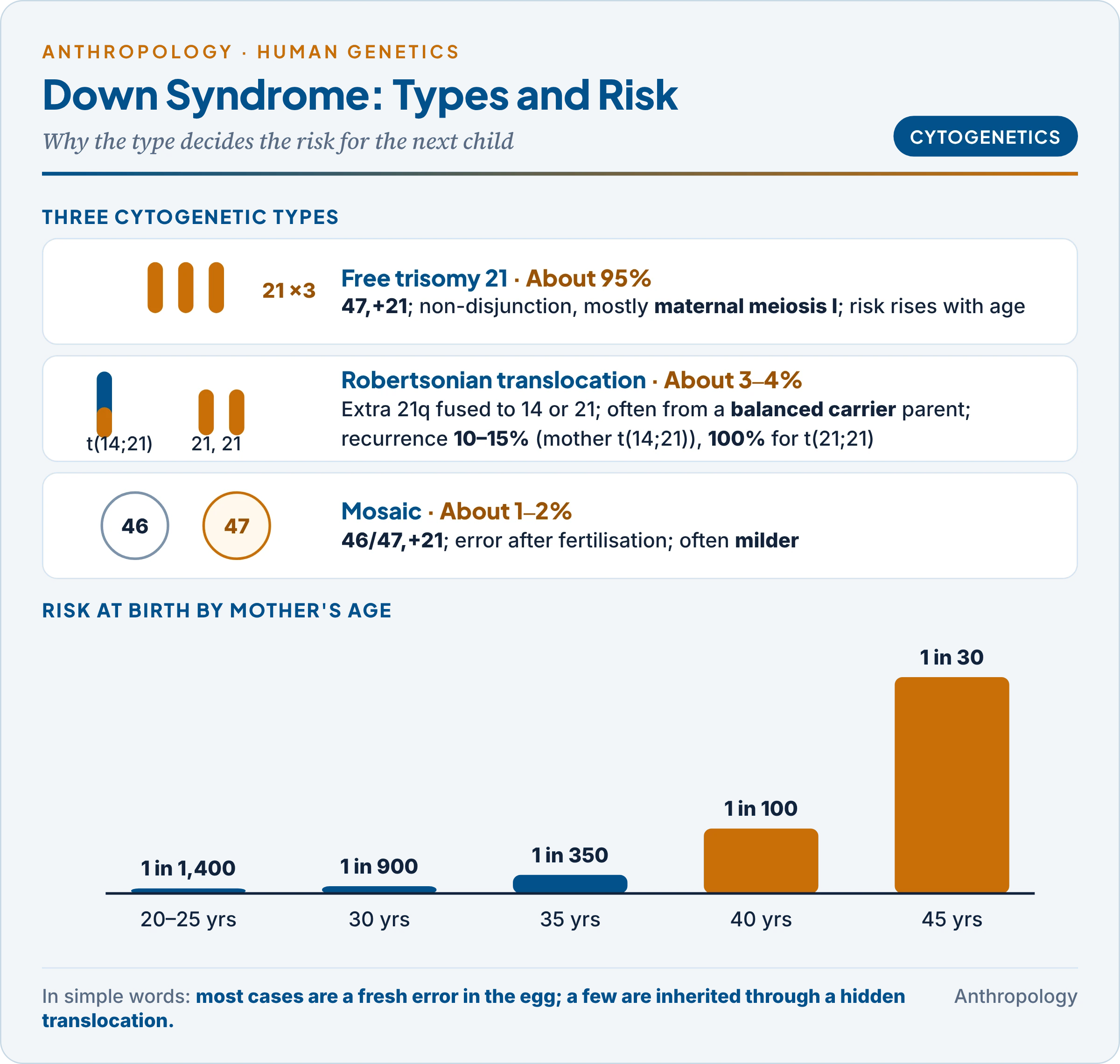
Three cytogenetic types. The type matters because it decides the risk to the next child.
| Type | Share | Mechanism | Recurrence |
|---|---|---|---|
| Free trisomy 21 (47,+21) | About 95% | Non-disjunction, mostly maternal meiosis I | Low; rises with maternal age |
| Robertsonian translocation (46,t(14;21) or t(21;21)) | About 3–4% | Extra 21q fused to chromosome 14 or 21; often inherited from a balanced carrier parent with 45 chromosomes | About 10–15% if the mother carries t(14;21), lower if the father; 100% for t(21;21) |
| Mosaic (46/47,+21) | About 1–2% | Mitotic error after fertilisation | Low; features often milder |
So when a child has translocation Down syndrome, the parents are karyotyped: a healthy carrier can have further affected children at any age.
| Mother’s age | Approximate risk at birth |
|---|---|
| 20–25 | 1 in 1,400 |
| 30 | 1 in 900 |
| 35 | 1 in 350 |
| 40 | 1 in 100 |
| 45 | 1 in 30 |
4.2 Patau, Edwards and cri-du-chat syndromes
The three rarer syndromes are compared in one table; all are diagnosed by karyotype and offered prenatal diagnosis in the next pregnancy.
| Feature | Patau | Edwards | Cri-du-chat |
|---|---|---|---|
| Karyotype | 47,+13 | 47,+18 | 46,5p− (deletion) |
| Described | Klaus Patau, 1960 | John H. Edwards, 1960 | Jérôme Lejeune, 1963 |
| Frequency | About 1 in 10,000–16,000 | About 1 in 5,000–6,000 | About 1 in 15,000–50,000 |
| Key signs | Holoprosencephaly (undivided forebrain), cleft lip and palate, small eyes, polydactyly | Clenched fists with overlapping fingers, rocker-bottom feet, small jaw, heart defects | High-pitched cat-like cry (abnormal larynx), microcephaly (small head), wide-set eyes, intellectual disability |
| Survival | Most die in the first year | About 90% die in the first year | Often survives to adulthood |
Why cri-du-chat differs: it is a structural error (a missing piece of 5p), not an extra chromosome, so the child lacks only some genes and can live long. About 10–15 per cent of cases come from a parent carrying a balanced translocation involving 5p, which is why parents are tested.
5. Structural Aberrations
In simple words
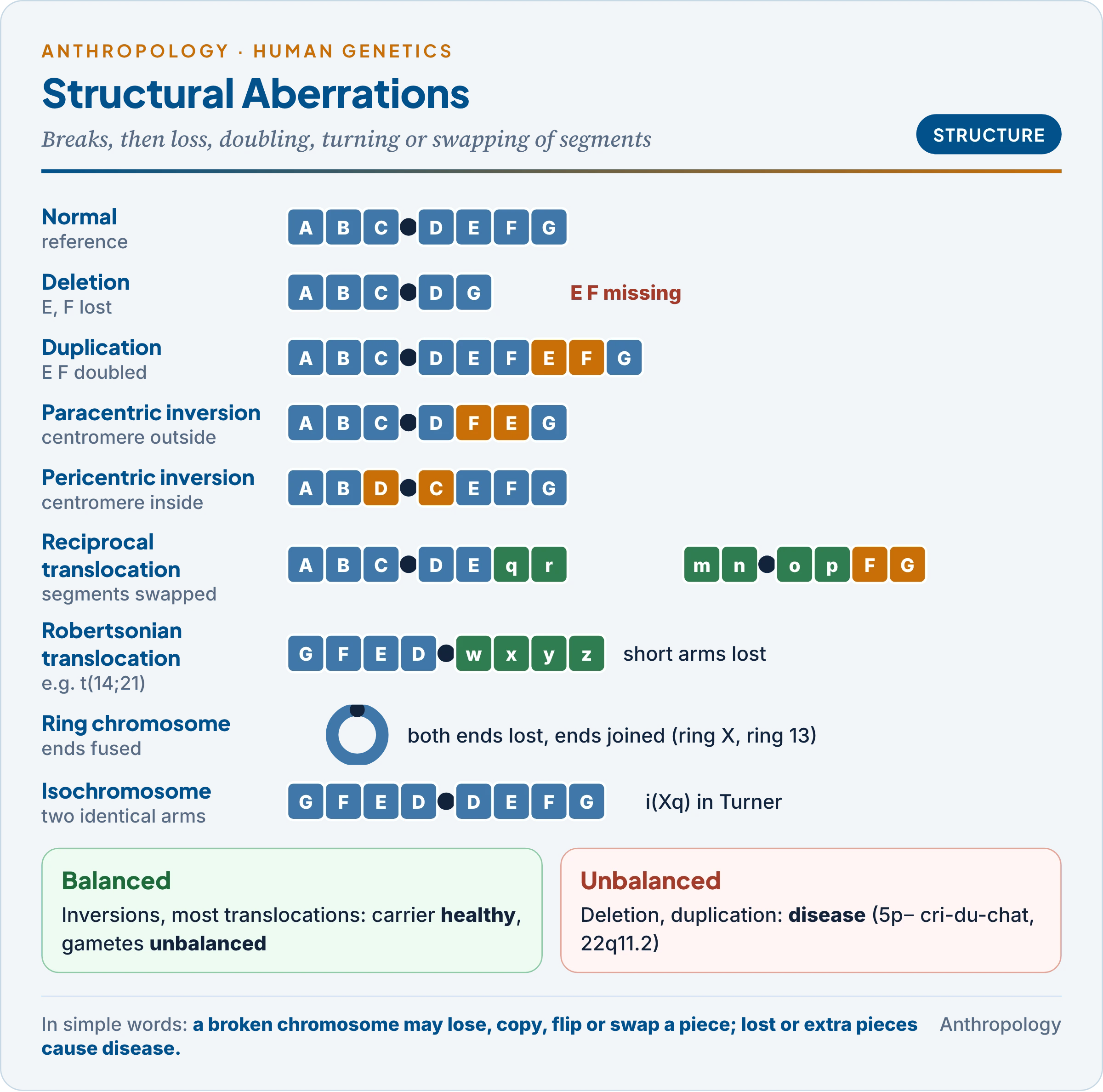
A structural aberration happens when a chromosome breaks and the pieces are lost, copied, turned round or joined to the wrong chromosome. If no genetic material is gained or lost, the person is usually healthy but may have children with problems; if material is gained or lost, a disorder results.
5.1 How breaks arise
- Spontaneous: faulty DNA repair and unequal crossing over between similar repeated sequences, which is why the same microdeletions recur in unrelated people.
- Induced: ionising radiation, chemical mutagens and some viruses.
- Balanced vs unbalanced: in a balanced rearrangement no material is lost (inversions, most reciprocal translocations): carriers are healthy but have unbalanced gametes, causing miscarriage or an affected child. An unbalanced change causes disease directly.
5.2 The main types
| Type | What happens | Example |
|---|---|---|
| Deletion | A segment is lost, terminal (end) or interstitial (middle); microdeletion if below microscope resolution | 5p− cri-du-chat; 22q11.2 DiGeorge syndrome; 15q11–q13 Prader-Willi and Angelman |
| Duplication | A segment is doubled, tandem (ABC-ABC) or reverse (ABC-CBA) | 17p12 duplication in Charcot-Marie-Tooth neuropathy; duplication also creates new gene families (globins) in evolution |
| Inversion | A segment turns 180°: paracentric (centromere outside) or pericentric (centromere inside) | inv(9), a harmless variant; pericentric inversions distinguish human and chimpanzee chromosomes |
| Reciprocal translocation | Two non-homologous chromosomes exchange segments | Philadelphia chromosome t(9;22) in chronic myeloid leukaemia; t(8;14) in Burkitt lymphoma |
| Robertsonian translocation | Two acrocentric chromosomes (13, 14, 15, 21, 22) fuse at the centromere; short arms lost | t(14;21) carrier with 45 chromosomes; about 1 in 1,000 people |
| Ring chromosome | Both ends break and join | Ring X in Turner; ring 13 |
| Isochromosome | The centromere divides crosswise, giving two identical arms | i(Xq) in Turner syndrome |
- Inversion loops: in an inversion carrier the paired chromosomes form a loop at meiosis; a crossover inside it gives dicentric and acentric products (paracentric) or duplicated and deleted products (pericentric), which is why carriers have more miscarriages.
- Translocation and cancer: the Philadelphia chromosome, found by Peter Nowell and David Hungerford (1960), joins BCR to the ABL gene, creating a fused gene that drives cell division: an activated oncogene (a gene whose overactivity causes cancer). The drug imatinib, which blocks the fused protein, was the first cancer treatment designed against a chromosomal change.
- Evolution: human chromosome 2 arose by the fusion of two ape chromosomes (great apes have 48), and several pericentric inversions separate human and chimpanzee chromosomes: structural change is also a source of evolution, not only disease.
6. Genetic Imprints in Human Disease
In simple words
For most genes it does not matter whether a copy came from the mother or the father. For a small group of genes it does: one parent’s copy is switched off before birth. If the working copy is lost, a disease appears, and which disease depends on which parent the loss came from.
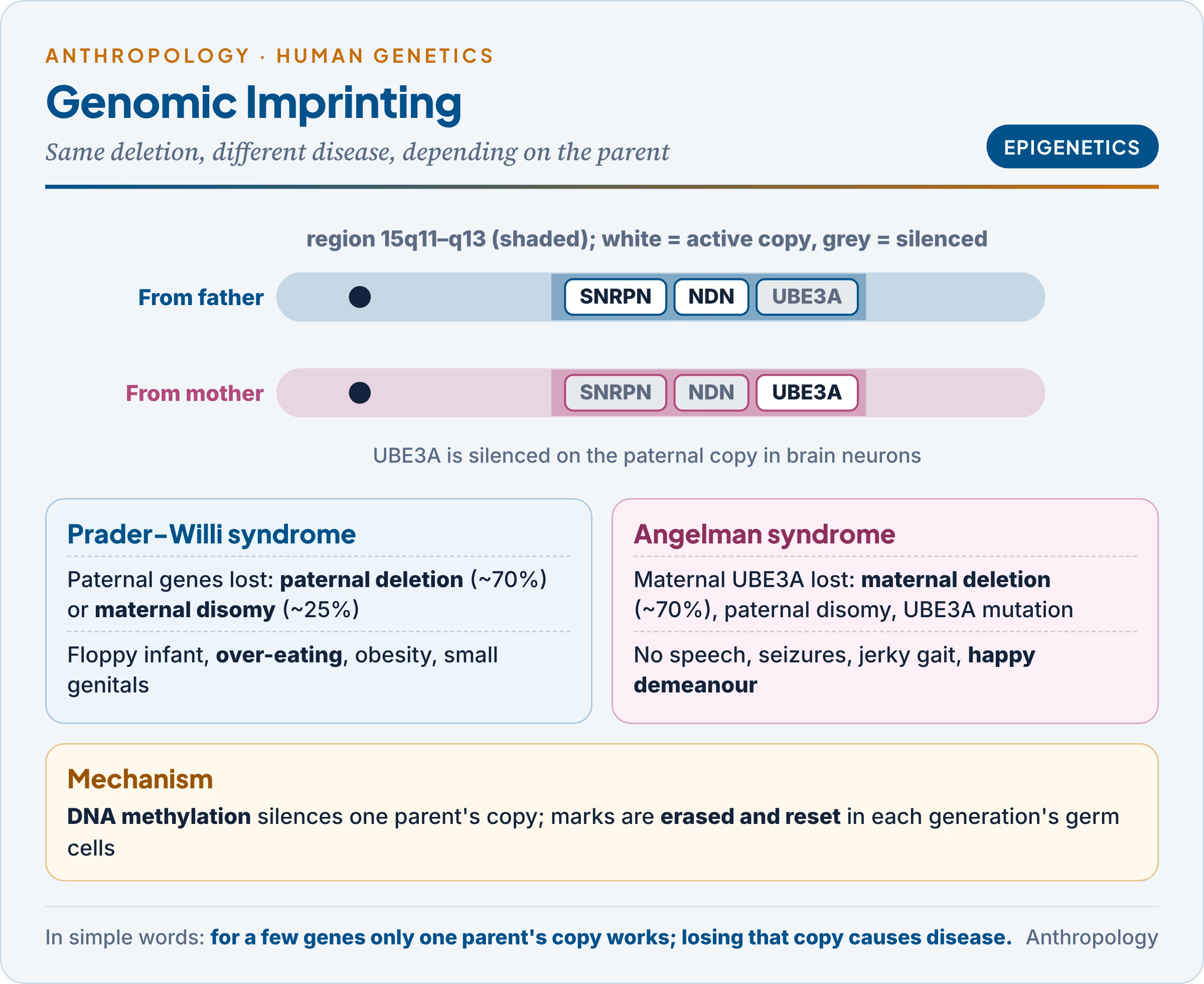
Two children both lack the same small piece of chromosome 15 (15q11–q13). One is floppy at birth, later eats compulsively, becomes obese and has small genitals: Prader-Willi syndrome. The other has severe intellectual disability, seizures, jerky movements and frequent laughter: Angelman syndrome. The only difference is that the first child’s deletion is on the chromosome from the father, the second’s on the one from the mother. This is genomic imprinting.
6.1 Concept and mechanism
- Mendel’s assumption broken: Mendel’s reciprocal crosses (tall female × dwarf male and the reverse) gave identical results, so classical genetics assumed parental equivalence. Imprinting is the exception: a gene’s expression depends on its parent of origin.
- Mechanism: DNA methylation (adding methyl groups to cytosine) and histone changes at an imprinting control region silence one parental copy. The mark is epigenetic: it changes gene activity, not the DNA sequence.
- Life cycle: marks are erased in the germ cells of each new generation and reset according to the sex of that individual, so a man passes paternal marks on all his chromosomes, including those from his mother.
- Scale: only about 100–200 of some 20,000 human genes are imprinted, and imprinting varies between species.
- Discovery: mouse embryos with two maternal or two paternal genomes failed to develop (McGrath and Solter; Surani, 1984). In humans a hydatidiform mole (two paternal sets) is placenta without embryo; an ovarian teratoma (two maternal sets) is tissue without placenta.
- Why imprinting exists (conflict theory): the American biologist David Haig argued that paternal genes favour drawing more maternal resources into the fetus (growth-promoting IGF2 is paternally expressed), while maternal genes restrain growth to spread resources over all her children.
6.2 Imprinting disorders
Each disorder arises in more than one way; what they share is the loss of the active parental copy.
| Disorder | Region | Usual mechanisms | Main features |
|---|---|---|---|
| Prader-Willi | 15q11–q13: paternal genes (SNRPN, NDN, MAGEL2) lost | Paternal deletion (about 70%); maternal uniparental disomy (both 15s from mother, about 25%); imprinting defect | Neonatal hypotonia, hyperphagia and obesity, hypogonadism, short stature |
| Angelman | 15q11–q13: maternal UBE3A lost in brain | Maternal deletion (about 70%); paternal disomy; UBE3A mutation; imprinting defect | Severe intellectual disability, absent speech, seizures, ataxia, happy demeanour |
| Beckwith–Wiedemann | 11p15: excess IGF2 | Paternal disomy or methylation change | Overgrowth, large tongue, Wilms tumour risk |
| Silver–Russell | 11p15 (or 7) | Loss of paternal methylation | Growth restriction, triangular face |
- Uniparental disomy (both copies of a chromosome from one parent) arises when a trisomic embryo loses the extra chromosome (“trisomy rescue“): a normal chromosome count can still cause an imprinting disease.
- Cancer: loss of imprinting of IGF2 occurs in Wilms tumour; reduced expression of the paternally expressed DIRAS3 is reported in many ovarian and breast cancers.
- Hereditary paraganglioma (glomus tumour, SDHD gene) appears almost only when the mutation comes from the father.
7. Genetic Screening
In simple words
Genetic screening means testing many people who have no symptoms to find those at high risk: carriers of a recessive gene, pregnancies with a chromosomal problem, or newborns with a treatable disorder. Those who screen positive are then given a definite diagnostic test.
7.1 Principles and types
Screening sorts people into higher and lower risk; diagnosis confirms. The WHO principles of Wilson and Jungner (1968) require an important condition with a known natural history, a valid, acceptable test, an effective treatment or choice, and justified cost. A test is judged by sensitivity (true cases detected) and specificity (unaffected correctly reassured).
| Type | Who is tested | Examples |
|---|---|---|
| Carrier (heterozygote) screening | Communities with high carrier frequency | Tay-Sachs among Ashkenazi Jews; thalassaemia in Cyprus, Sardinia and India; sickle cell in tribal India (Unit 9.1) |
| Prenatal screening | Pregnant women | First-trimester combined test (nuchal translucency on ultrasound plus PAPP-A and hCG in serum), quadruple test, NIPT; maternal serum alpha-fetoprotein for neural tube defects |
| Prenatal diagnosis | Screen-positive or high-risk pregnancies | Chorionic villus sampling (10–13 weeks), amniocentesis (15–20 weeks), cordocentesis (umbilical cord blood); karyotype, FISH or DNA test |
| Preimplantation genetic testing | Couples using IVF | Testing embryos before transfer |
| Newborn screening | All newborns | Heel-prick blood spot (Guthrie, 1963, for phenylketonuria); congenital hypothyroidism, G6PD deficiency, galactosaemia |
| Predictive testing | Adults with a family history | BRCA1/BRCA2 for breast and ovarian cancer; Huntington disease |
- Why carrier screening works best in defined communities: recessive disorders are common where endogamy keeps carriers marrying carriers. The Cyprus programme, backed from 1983 by a premarital certificate, reduced thalassaemia-major births to near zero. Reich and colleagues (2009) predicted that India’s many endogamous groups would each carry their own excess of recessive disease (Unit 9.5).
- Why newborn screening pays: a diet started early prevents disability in phenylketonuria, and a daily thyroxine tablet does so in congenital hypothyroidism.
7.2 Indian framework
- PCPNDT Act, 1994 (Pre-Conception and Pre-Natal Diagnostic Techniques (Prohibition of Sex Selection) Act; in force 1996, renamed and strengthened 2003):
- s.3: only registered genetic counselling centres, laboratories and clinics may conduct prenatal diagnosis; s.3A prohibits sex selection.
- s.4(2): prenatal tests are allowed only to detect chromosomal abnormalities, genetic metabolic diseases, haemoglobinopathies, sex-linked genetic diseases and congenital anomalies.
- s.4(3): a test needs one of these recorded conditions: mother above 35 years, two or more spontaneous abortions or fetal loss, exposure to teratogens (drugs, radiation, infection, chemicals), or a family history of mental retardation, physical deformity or genetic disease.
- s.5: written consent is required, and the sex of the fetus may not be communicated by words, signs or any other method.
- Why the law exists: the same amniocentesis that detects Down syndrome also reveals fetal sex, and its misuse for sex-selective abortion lowered India’s child sex ratio to 918 girls per 1,000 boys (Census 2011). The Act keeps diagnosis available while blocking sex determination.
- MTP (Amendment) Act, 2021: termination beyond 24 weeks is permitted for substantial fetal abnormalities diagnosed by a Medical Board, which makes late prenatal diagnosis meaningful.
- Rashtriya Bal Swasthya Karyakram (2013) screens children for the “4 Ds” (defects at birth, diseases, deficiencies, developmental delays), including Down syndrome; universal newborn biochemical screening exists only in some states.
8. Genetic Counselling
In simple words
Genetic counselling helps a family understand a genetic condition: what it is, how it is inherited, the chance that it will happen again, and the choices available. The counsellor explains and supports; the family decides.
8.1 Definition and aims
The term was coined by the American geneticist Sheldon Reed (1947). The American Society of Human Genetics (committee chaired by F. Clarke Fraser, 1975) defined it as “a communication process which deals with the human problems associated with the occurrence, or the risk of occurrence, of a genetic disorder in a family.” Its aims are to help the family understand the diagnosis, the inheritance and recurrence risk, the options, and to make and adjust to a decision.
8.2 Steps
- Referral and intake: an affected child, recurrent miscarriage, infertility, consanguinity, an abnormal screen or disputed paternity.
- Pedigree: at least three generations, recording consanguinity, miscarriages and deaths (Unit 9.1).
- Diagnosis: examination and tests (karyotype, FISH, biochemical or DNA); without a firm diagnosis the risk figures may be wrong.
- Risk assessment: Mendelian (both parents carriers: 1 in 4 affected; dominant: 1 in 2), empirical (observed recurrence for multifactorial conditions such as cleft lip, about 3–4 per cent after one affected child) or Bayesian (combining prior risk with test results).
- Communication of options: prenatal diagnosis, preimplantation testing, donor gametes, adoption, or accepting the risk.
- Decision and support: non-directive counselling (the counsellor does not tell the family what to choose); psychosocial support and referral to support groups.
- Follow-up: testing of relatives (cascade screening), and updates as knowledge changes.
Why non-directiveness matters: genetic counselling grew in the shadow of eugenics (Unit 12), when states decided who should reproduce. Respect for autonomy separates counselling from eugenic control.
8.3 Problems
- Mistaken paternity revealed by testing; confidentiality versus the relatives’ interest in knowing their own risk.
- Discrimination by employers and insurers: the US Genetic Information Nondiscrimination Act (2008) is one response.
- Uncertainty: distinguishing a harmless polymorphism from a disease variant (a “variant of uncertain significance”).
- Cultural context: in South India, where cousin and uncle–niece marriage is a valued kinship preference, counselling must give the higher recessive risk without condemning the custom.
- Misuse: sex selection; pressure to terminate; the lingering eugenic idea that carriers should not marry. Recessive alleles hide in heterozygotes, so even preventing all affected births lowers their frequency only slowly.
8.4 Gene therapy
- Somatic therapy changes the patient’s own cells; germ-line therapy (embryo editing) would change descendants and is banned in most countries; the gene-edited twins born in China (He Jiankui, 2018) were widely condemned.
- Milestones: first approved trial for ADA deficiency (US, 1990); the CRISPR therapy Casgevy for sickle-cell disease and beta-thalassaemia (UK and US, 2023).
9. Human DNA Profiling
In simple words
DNA profiling identifies a person by looking at a few places in the DNA where people differ greatly in the number of times a short sequence is repeated. It is used to solve crimes, settle parentage and identify the dead.
The principle, PCR and the logic of paternity testing were explained in Unit 9.1 (§8.4): a child carries one allele at each locus from each parent, so an alleged father lacking the child’s paternal allele at several loci is excluded. Here the methods and their limits are compared.
| Method | Marker | Strength | Limit |
|---|---|---|---|
| RFLP–VNTR (Alec Jeffreys, 1984) | Minisatellites (10–100 base repeats), cut by restriction enzymes, separated by electrophoresis, Southern blot, radioactive probe | Highly variable | Needs much fresh DNA; slow |
| PCR–STR (since the 1990s) | Microsatellites (2–6 base repeats) at 15–24 standard loci | Works on traces and old samples; standard databases | Mixtures and contamination |
| Y-STR | Y chromosome | Male line; sexual-assault mixtures | Shared by all paternal relatives |
| Mitochondrial DNA | Maternal line | Hair shafts, bones | Shared by all maternal relatives |
- Probability, not certainty: a match is reported as a random match probability from allele frequencies in a reference population; because frequencies differ between populations, the database matters, and grouping people by “race” for it raises the questions of Unit 9.5.
- India: DNA fingerprinting was developed at CCMB, Hyderabad, by Lalji Singh (1988); the Centre for DNA Fingerprinting and Diagnostics (CDFD) followed. The DNA Technology (Use and Application) Regulation Bill, 2019, which proposed a national DNA data bank, was withdrawn on 24 July 2023, the government stating that the Criminal Procedure (Identification) Act, 2022, which allows biological samples to be taken as “measurements”, covers most of it. Critics note the loss of the Bill’s safeguards: accreditation of laboratories, quality standards and rules on deleting profiles.
10. Gene Mapping and Genome Study
In simple words
Gene mapping finds where a gene sits on a chromosome. Genome study reads the whole DNA sequence, about three billion letters. Together they let doctors find disease genes, understand human variation and trace our ancestry.
From 1979 the American psychologist Nancy Wexler collected blood from a huge family around Lake Maracaibo, Venezuela, in which Huntington disease was common. By following DNA markers through this pedigree, James Gusella and colleagues (1983) placed the gene on chromosome 4; the gene itself was isolated in 1993. This is gene mapping by linkage: a marker that travels with the disease through a family must lie close to the gene.
10.1 Kinds of maps and methods
| Map | Unit | Built by |
|---|---|---|
| Genetic (linkage) map | Centimorgan (1% recombination) | Following markers through families; LOD score (likelihood of linkage, Newton Morton, 1955) |
| Cytogenetic map | Chromosome bands | Banding, FISH, somatic-cell hybrids (human–mouse cells that lose human chromosomes) |
| Physical map | Base pairs | Restriction fragments, cloned overlapping fragments (contigs) |
| Sequence map | Individual bases | DNA sequencing |
- Genome-wide association studies (GWAS): instead of following families, a GWAS compares hundreds of thousands of SNPs in thousands of patients and controls and looks for variants more common in patients (significance threshold p < 5 × 10⁻⁸). The first notable success found a complement factor H variant in age-related macular degeneration (2005); the Wellcome Trust Case Control Consortium (2007) studied seven common diseases at once.
- What GWAS taught: common diseases (diabetes, heart disease, schizophrenia) are polygenic, each variant adding a tiny risk, hence polygenic risk scores. Limits: hits explain little of the heritability (“missing heritability“), and most samples are European, so results transfer poorly to Indians.
10.2 The Human Genome Project
- Origin: the US Department of Energy sought better ways to detect mutations in Hiroshima and Nagasaki survivors (Alta, Utah, 1984); a National Research Council report (1988) proposed sequencing the genome with the National Institutes of Health; HUGO (1988) coordinated internationally.
- Course: launched October 1990 as a 15-year, US$3 billion effort led first by James Watson; joined by the UK (Sanger Centre), France, Germany, Japan and China; a private rival, Celera Genomics (Craig Venter), sped it up. The draft was announced on 26 June 2000 and published in February 2001; the project was declared complete in April 2003.
- Goals: map and sequence the 3 billion base pairs; identify all genes; store data publicly; develop analysis tools; address ethical, legal and social implications (ELSI, set up 1990).
- Findings: only about 20,000 protein-coding genes (earlier estimates were 100,000); under 2 per cent of the genome codes for protein; any two people are about 99.9 per cent identical.
- Completion: the Telomere-to-Telomere consortium filled the last gaps (2022), and a pangenome of 47 people from diverse ancestries (2023) replaced the single mosaic reference.
- Criticism: “big science” diverting funds from small projects; most of the genome dismissed as junk (much is now known to regulate genes); privacy, genetic discrimination and patenting; over-promised cures.
10.3 Genome study in India and anthropology
- IndiGen (CSIR, 2019): whole genomes of 1,029 Indians (1,008 announced in October 2019), with genome cards for clinically relevant variants.
- GenomeIndia (DBT): 10,074 genomes from 83 populations, data opened in January 2025 (Unit 9.1).
- Population history: genome data revealed the ANI–ASI mixture behind most Indian groups (Unit 9.5) and Neanderthal ancestry in all non-Africans (Svante Pääbo; Nobel Prize 2022).
- Applications: rare-disease diagnosis, carrier screening, pharmacogenomics (choosing drugs by genotype, such as testing for HLA-B*15:02 before the anti-epileptic carbamazepine), cancer genomics, forensic identification and tracing migrations.
Key Terms
| Term | Meaning in simple words |
|---|---|
| Karyotype | A picture of a cell’s chromosomes arranged in pairs |
| Aneuploidy | One chromosome extra or missing |
| Polyploidy | One or more whole extra sets of chromosomes |
| Non-disjunction | Failure of chromosomes to separate when gametes form |
| Mosaicism | Two cell lines with different chromosomes in one person |
| Barr body | The switched-off X chromosome seen in female cells |
| SRY | The gene on Y that starts male development |
| Robertsonian translocation | Two acrocentric chromosomes joined at the centromere |
| Microdeletion | A deletion too small to see under the microscope |
| Genomic imprinting | A gene’s activity depending on which parent it came from |
| Uniparental disomy | Both copies of a chromosome inherited from one parent |
| Carrier screening | Testing healthy people for one copy of a recessive gene |
| NIPT | A blood test of the mother that screens the fetus’s DNA |
| Non-directive counselling | Counselling that informs but leaves the decision to the family |
| STR | A short DNA sequence repeated a variable number of times, used in profiling |
| GWAS | A study scanning the whole genome for variants linked to a disease |
Contemporary Developments
- DNA Bill withdrawn (2023): the DNA Technology Regulation Bill, 2019 was withdrawn on 24 July 2023 because the Criminal Procedure (Identification) Act, 2022 already allows biological samples to be taken; its laboratory-accreditation and data-deletion safeguards lapsed with it.
- Gene editing reaches the clinic (2023): Casgevy, the first CRISPR therapy, was approved for sickle-cell disease and beta-thalassaemia, two disorders India screens for on a large scale.
- Complete and diverse references (2022–23): the gapless T2T genome and the first pangenome improved the detection of structural variants that older maps missed.
- GenomeIndia (2025): 10,074 Indian genomes from 83 populations opened to researchers, allowing community-specific carrier screening and reducing the European bias of GWAS.
Diagrams to Master
- Topic map: chromosomes and methods → numerical, sex-chromosome, autosomal, structural → imprinting → screening, counselling, profiling, mapping.
- Non-disjunction: meiosis I (all four gametes abnormal: two n+1, two n−1) vs meiosis II (one n+1, one n−1, two normal); trisomy and monosomy after fertilisation.
- Sex-chromosome aneuploidies: 47,XXY / 45,X / 47,XXX / 47,XYY with Barr bodies (X − 1), frequency and features.
- Down syndrome types: free trisomy 95%, t(14;21) 3–4%, mosaic 1–2%; maternal-age curve (1 in 350 at 35, 1 in 100 at 40).
- Structural aberrations: lettered chromosome (A–G) showing deletion, duplication, inversion (para/peri), reciprocal and Robertsonian translocation, ring, isochromosome.
- Imprinting at 15q11–q13: paternal genes (SNRPN, NDN) vs maternal UBE3A; paternal loss → Prader-Willi, maternal loss → Angelman.
Revision Grid
| Scholar | Work (year) | Key idea |
|---|---|---|
| John Langdon Down | 1866 | Described the syndrome; racial label “mongolism” |
| Henry Turner | 1938 | Turner syndrome described |
| Harry Klinefelter | 1942 | Klinefelter syndrome described |
| Jérôme Lejeune | 1959; 1963 | Trisomy 21; cri-du-chat (5p−) |
| Ford; Jacobs and Strong | 1959 | 45,X and 47,XXY |
| Patau; Edwards | 1960 | Trisomy 13; trisomy 18 |
| Mary Lyon | 1961 | X-inactivation |
| Patricia Jacobs | 1965 | XYY survey; later “criminal” myth refuted |
| Wilson and Jungner | 1968 | WHO screening principles |
| Fraser (ASHG) | 1975 | Definition of genetic counselling |
| Gusella and colleagues | 1983 | Huntington gene mapped to chromosome 4 |
| McGrath and Solter; Surani | 1984 | Imprinting shown in mouse embryos |
| Alec Jeffreys | 1984 | DNA fingerprinting |
| Haig and Westoby | 1989 | Parental conflict theory of imprinting |
| Human Genome Project | 1990–2003 | Human genome sequenced; about 20,000 genes |
Previous Year Questions
Unit 9.4: Chromosomes and chromosomal aberrations in man, methodology
- 2026Numerical chromosomal aberrations can be due to abnormalities in autosomes as well as sex chromosomes. Elaborate in terms of the effects these have on human body, giving suitable examples.
- 2025‘Genome-wide Disease Association Studies (GWAS) advanced our understanding of health and disease.’ Discuss.
- 2024What is meant by karyotype? How does its analysis help in diagnosis of the chromosomal aberrations in man?
- 2024What is genetic counselling? Briefly discuss various steps involved in it.
- 2023Describe the causes of structural abnormalities of chromosomes with suitable examples.
- 2023Describe the practical applications of DNA technology in the current scenario.
- 2022Write notes on: Genetic imprinting in human diseases.
- 2021“Chromosomal aberrations can play havoc with the human body and mind.” Explain with suitable examples.
- 2020How may numerical aberrations in sex chromosomes lead to genetic disorders ?
- 2020What are the applications of human genomic research in human welfare?
- 2018Write a note in 150 words on Categorize Genes that influence Human Survival.
- 2018Describe the mechanisms for structural anomalies of autosomes with diagrams.
- 2016Explain the significance of screening and counselling for genetic disorders.
- 2015Write a note in 150 words on Down’s Syndrome.
- 2015Discuss chromosomal aberrations in man illustrating with examples.
- 2014Write a note in 150 words on Genetic Counselling.
- 2014Describe Turner and Klinefelter Syndromes in humans.
- 2012Write a note in 150 words on Genetic Counselling.
- 2012Discuss the chromosomal aberrations and manifestations of Klinefelter and Turner syndromes.
- 2010Write a note in 150 words on Oncogenes.
- 2010Chromosomal deletions and numerical fluctuations may lead to gross abnormalities in man. Discuss with the help of suitable examples.
- 2007Write a note on Genome Study.
- 2007Discuss different types of sex chromosomal aberrations.
- 2006What is Genetic Counselling? Discuss its relevance in the present day context.
- 2003Write a note on Klinefelter syndrome.
- 2002Write a note on Genetic Counselling.
- 2001Discuss the relevance of human DNA Profiling and Gene Mapping in the prevention and cure of diseases.
- 1998Write a note on Genetic counselling.
- 1995Write a note on Gene therapy.
- 1987Write a note on Taurodontism.
Related questions from other units
- 2026Unit 12: In what manner is our understanding of serogenetics and cytogenetics useful in reproductive biology? What are the major ethical issues involved in reproductive technology?
- 2022Unit 9.1: Write notes on: Pedigree analysis in genetic counselling.
- 2021Unit 12: How are the cases of disputed paternity solved ? Discuss the recent techniques.
- 2020Unit 12: Write notes on the Ethics and Genetic engineering in about 150 words.
- 2019Unit 12: “Applied human genetics has come to touch every sphere of human life.” Discuss in light of recent advances in molecular anthropology.
- 2015Unit 12: Write a note in 150 words on DNA technology in Medicine.
- 2011Unit 12: Explain the application of anthropological knowledge in genetic counseling, forensic science, sports and nutrition.
- 2001Unit 12: Evaluate the role of Serogenetics and Cytogenetics in reproductive biology.
- 1996Unit 12: Discuss the concepts of eugenics and euphenics and their potential applications to human welfare.
- 1995Unit 9.1: Discuss the genetical and clinical aspects of the anomalies of sex chromosomes in man with special reference to the associated mosaic constitutions.