

1.7 The biological basis of life: The Cell, DNA structure and Replication, Protein Synthesis, Gene, Mutation, Chromosomes and Cell Division
Official Syllabus: Paper I, Unit 1.7
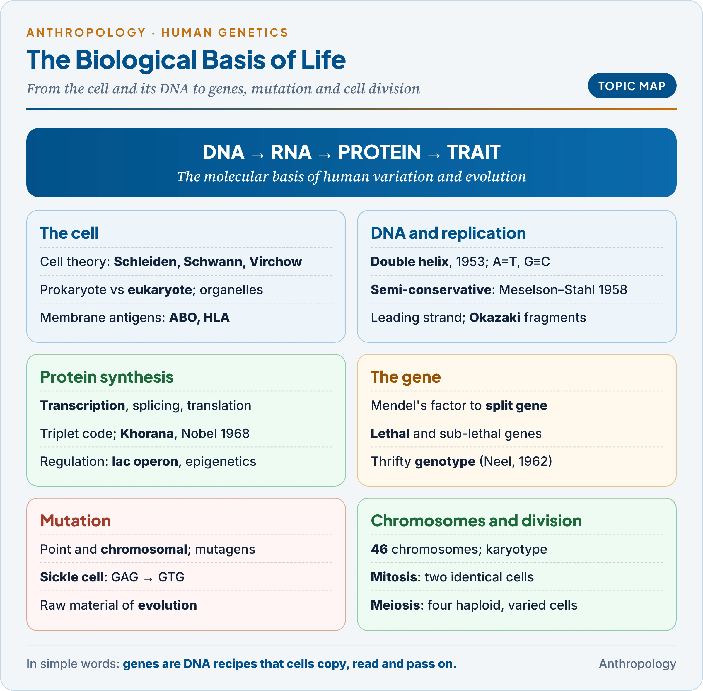
Physical anthropology studies human variation and evolution, and both rest on the molecules of the cell. The sickle-cell gene of India’s tribal belt, the mitochondrial DNA that traces our African origin, the ancient DNA of Neanderthals (Unit 1.6c) and the chromosomal disorders of Unit 9.4 all make sense only once the cell, DNA, genes and cell division are understood. The forces that change gene frequencies are in Unit 1.4b (in brief: mutation supplies new variation, and selection, drift and gene flow sort it); human genetic methods are in Unit 9.
1. The Cell
In simple words
The cell is the smallest unit that can live on its own. Every living thing is made of one or more cells, and every cell comes from an earlier cell. Inside our cells a nucleus holds the DNA, and small working parts called organelles make proteins, release energy and package materials.
1.1 Cell theory
In 1665 the English scientist Robert Hooke saw the walls of dead cork and called them “cells”; Antonie van Leeuwenhoek saw living cells in the 1670s; Robert Brown described the nucleus in 1831.
- Cell theory: the German botanist Matthias Schleiden (1838) and the German physiologist Theodor Schwann (1839) proposed that all plants and animals are made of cells; the German pathologist Rudolf Virchow (1855) added that every cell arises from a pre-existing cell (omnis cellula e cellula). The theory makes the cell the unit of structure, function and inheritance: growth, repair and reproduction are all cell division.
- Scale: a human body has roughly 30–37 trillion cells.
1.2 Prokaryotic and eukaryotic cells
Cells come in two basic designs; the key difference is the nucleus and membrane-bound organelles.
| Feature | Prokaryotic cell | Eukaryotic cell |
|---|---|---|
| Examples | Bacteria, blue-green algae, mycoplasma | Protists, fungi, plants, animals |
| Nucleus | None; DNA lies free in the cytoplasm | True nucleus with a double envelope |
| DNA | One circular chromosome, plus plasmids | Several linear chromosomes on histones |
| Organelles | None bound by membranes | Mitochondria, ER, Golgi, lysosomes |
| Ribosomes | 70S | 80S (70S in mitochondria) |
The 70S ribosomes and circular DNA inside mitochondria betray their past: in the endosymbiotic theory of the American biologist Lynn Margulis (1967), mitochondria descend from engulfed bacteria.
1.3 The parts of a eukaryotic cell
The table lists the main organelles; the last column shows why each matters to an anthropologist.
| Part | Function | Anthropological relevance |
|---|---|---|
| Plasma membrane (fluid mosaic model: Seymour Singer and Garth Nicolson, 1972) | Phospholipid bilayer with proteins; controls entry and exit | Carries the ABO and HLA antigens used as genetic markers (Unit 9.6) |
| Nucleus, with nucleolus | Stores DNA; makes RNA; nucleolus makes ribosomes | Source of nuclear DNA |
| Ribosomes; endoplasmic reticulum | Ribosomes make proteins; rough ER carries ribosomes, smooth ER makes lipids and steroid hormones | Translation (§4); hormones of growth (Unit 10) |
| Golgi apparatus (Camillo Golgi, 1898) | Modifies, packages and dispatches proteins; adds sugars | Builds the sugar chains of the ABO antigens |
| Lysosomes (Christian de Duve, 1955) | Digestive enzymes | Missing enzymes cause storage diseases such as Tay-Sachs |
| Mitochondria | Aerobic respiration: make energy as ATP | Own mtDNA, inherited from the mother (§9) |
| Centrosome | Pair of centrioles; organises the spindle | Errors cause aneuploidy (§8) |
2. DNA: The Genetic Material and Its Structure
In simple words
DNA is a very long molecule shaped like a twisted ladder. Its rungs are pairs of four chemical “letters”, A, T, G and C, and the order of these letters is the genetic message. Because A always pairs with T and G with C, each side of the ladder carries the information needed to rebuild the other.
2.1 How DNA was shown to be the genetic material
Each experiment answered one question, which is why they are taught in sequence.
| Experiment | Year | What was done | What it proved |
|---|---|---|---|
| Frederick Griffith | 1928 | Heat-killed virulent pneumonia bacteria transformed harmless live ones | A “transforming principle” carries heredity |
| Oswald Avery, Colin MacLeod and Maclyn McCarty | 1944 | Destroyed each type of molecule in turn; only destroying DNA stopped transformation | The principle is DNA |
| Alfred Hershey and Martha Chase | 1952 | Labelled virus DNA and protein; only DNA entered bacteria | Viral genes are DNA, not protein |
| Erwin Chargaff | 1950 | Measured bases in many species | A = T and G = C |
2.2 The double helix
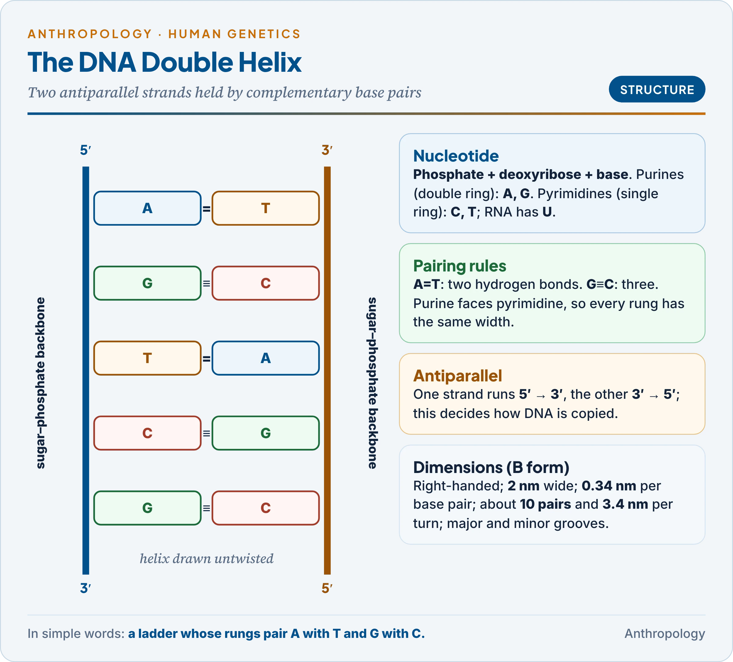
In April 1953 the American James Watson and the British Francis Crick proposed the double helix in Nature, using X-ray photographs by Rosalind Franklin (notably “Photo 51”, 1952) and Maurice Wilkins, and Chargaff’s rules.
- Building block: a nucleotide = phosphate + deoxyribose sugar + base. Bases are purines (double ring: adenine, guanine) or pyrimidines (single ring: cytosine, thymine; RNA has uracil instead of thymine).
- Backbone: sugars and phosphates joined by phosphodiester bonds form the rails; bases point inwards.
- Base pairing: A pairs with T by two hydrogen bonds, G with C by three. A purine always faces a pyrimidine, so every rung has the same width (helix about 2 nm across). The strands are therefore complementary: one fixes the sequence of the other.
- Antiparallel strands: one runs 5′ to 3′ (named after the carbon atoms of the sugar), the other 3′ to 5′, which decides how DNA is copied (§3).
- Dimensions: a right-handed helix with about 10 base pairs per turn, 0.34 nm between pairs and 3.4 nm per turn, with a major and a minor groove in which proteins read the bases.
- Why the structure explains heredity: each strand is a template for copying, the base sequence stores information, and a rare mispairing explains mutation.
2.3 DNA and RNA compared
The differences explain why DNA stores information and RNA works with it.
| Feature | DNA | RNA |
|---|---|---|
| Sugar | Deoxyribose | Ribose |
| Bases | A, G, C, T | A, G, C, U |
| Strands | Double helix | Usually single |
| Stability | Stable | Short-lived (most mRNA) |
| Role | Stores information | Carries and uses it: mRNA, tRNA, rRNA |
DNA’s double strand makes it chemically stable, which is why bones yield ancient genomes. The human genome has about 3.1 billion base pairs per set of 23 chromosomes and about 20,000 protein-coding genes; only about 1.5 per cent codes for protein, and the DNA of one cell, stretched out, would be about 2 m long.
3. DNA Replication
In simple words
Before a cell divides it copies all its DNA. The two strands unzip, and each old strand serves as a pattern for a new partner. So each new DNA molecule keeps one old strand and gets one new one, which makes copying very accurate.
3.1 Semi-conservative replication
Watson and Crick predicted that each daughter molecule would contain one old and one new strand: semi-conservative replication. The alternatives were conservative (the old helix stays whole beside a wholly new one) and dispersive (old and new pieces mixed).
- The proof: in 1958 Matthew Meselson and Franklin Stahl grew E. coli on heavy nitrogen (¹⁵N), moved it to normal nitrogen (¹⁴N) and separated DNA by density. After one generation all DNA was of intermediate density, ruling out the conservative model; after two, half was intermediate and half light, ruling out the dispersive model (Worked Example 2).
- In chromosomes: J. Herbert Taylor (1957) showed the same in broad-bean root cells using radioactive thymidine.
3.2 The machinery of replication
Copying happens in the S phase of the cell cycle (§8), from many origins of replication (one in E. coli; tens of thousands in a human cell).
| Enzyme or protein | What it does |
|---|---|
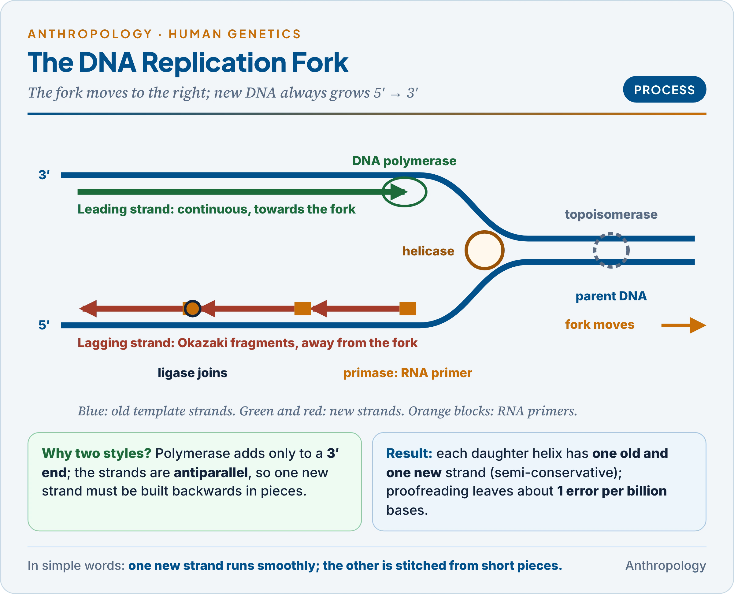
| Helicase | Breaks hydrogen bonds and opens the replication fork |
| Topoisomerase | Relieves the twisting ahead of the fork |
| Primase | Lays a short RNA primer, since DNA polymerase cannot start a chain |
| DNA polymerase | Adds nucleotides only to a free 3′ end, so builds 5′ to 3′; also proofreads |
| DNA ligase | Seals the pieces into one strand |
Leading and lagging strands. Because the strands are antiparallel and polymerase works only 5′ to 3′, one new strand (the leading strand) grows continuously towards the fork, while the other (the lagging strand) is made in short pieces, the Okazaki fragments (Reiji and Tsuneko Okazaki, 1968; 1,000–2,000 nucleotides in bacteria, 100–200 in eukaryotes), later joined by ligase.
- Accuracy: proofreading and mismatch repair cut errors to about one in a billion bases; the leftover errors are spontaneous mutations, about 60–70 new in each child (Unit 1.4b).
- Telomeres: the lagging strand cannot be finished at the tip of a linear chromosome, so ends shorten with each division. They are protected by telomeres (TTAGGG repeats), which telomerase extends in germ and stem cells (Elizabeth Blackburn, Carol Greider and Jack Szostak, Nobel Prize 2009). In most body cells telomeres shorten until division stops, part of the explanation of the Hayflick limit (about 50 divisions) and of ageing (Unit 10b).
4. Protein Synthesis and Gene Expression
In simple words
A gene is a stretch of DNA with the recipe for a protein. The cell first copies the recipe into a messenger molecule, RNA, which leaves the nucleus. Ribosomes then read the message three letters at a time and join amino acids in the right order. Cells switch genes on and off, so each cell makes only the proteins it needs.
4.1 From gene to protein
Proteins do the body’s work, and their link to genes was first seen in a human disease. In 1902 the English physician Archibald Garrod noticed that alkaptonuria (black urine) ran in families, often among children of cousin marriages, and behaved as a Mendelian recessive; he called such disorders “inborn errors of metabolism” (Croonian lectures, 1908): a missing enzyme caused by a faulty gene. George Beadle and Edward Tatum (1941) proved the link in the mould Neurospora: “one gene, one enzyme”, later refined to one gene, one polypeptide (one protein chain).
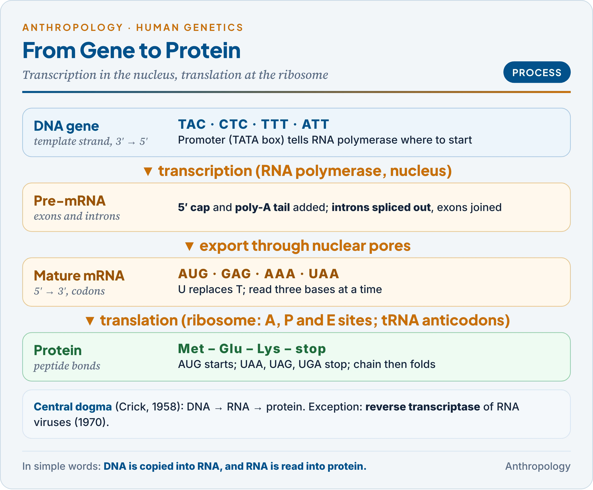
- The central dogma (Francis Crick, 1958): information flows DNA → RNA → protein. Howard Temin and David Baltimore (1970) found the exception, reverse transcriptase, by which RNA viruses such as HIV copy RNA into DNA.
4.2 Transcription: DNA to RNA
- How: in the nucleus, RNA polymerase binds the promoter, a control sequence upstream of the gene (often with a TATA box), unwinds the DNA and reads the template strand 3′ to 5′, building messenger RNA (mRNA) 5′ to 3′ with U opposite A. The mRNA thus matches the other, coding strand, with U for T.
- Processing: the pre-mRNA gets a protective 5′ cap and poly-A tail, and splicing removes non-coding introns and joins exons (split genes, discovered by Phillip Sharp and Richard Roberts, 1977); alternative splicing lets one gene make several proteins.
- Other RNAs: tRNA carries amino acids; rRNA forms the ribosome; microRNAs regulate genes.
4.3 The genetic code
The message is read in codons of three bases: two bases give only 16 combinations, too few for 20 amino acids, while three give 64 (Worked Example 3).
- Deciphering: Marshall Nirenberg and Heinrich Matthaei (1961) found that synthetic RNA of pure U makes a chain of phenylalanine (UUU = Phe); the Indian-born American chemist Har Gobind Khorana decoded the rest with repeating sequences, and Robert Holley solved tRNA structure (Nobel Prize, 1968).
- Properties:
- Triplet, read from a fixed start; AUG (methionine) starts, UAA, UAG and UGA stop.
- Degenerate: most amino acids have several codons, usually differing in the third base (Crick’s wobble hypothesis, 1966), so many base changes are silent.
- Unambiguous: each codon means one amino acid.
- Non-overlapping and comma-less, so an inserted or deleted base shifts the whole reading frame (§6).
- Nearly universal, strong evidence of the common origin of life; mitochondria are an exception (UGA = tryptophan).
4.4 Translation: RNA to protein
- Activation: each amino acid is joined to its own tRNA by an aminoacyl-tRNA synthetase, using ATP; the tRNA’s anticodon pairs with the codon.
- Initiation: the small ribosomal subunit binds the mRNA at the AUG start codon; the tRNA carrying methionine pairs with it; the large subunit joins.
- Elongation: the ribosome has three sites: A (arriving tRNA), P (holding the growing chain) and E (exit). A peptide bond joins the chain to each new amino acid (catalysed by the ribosome’s own rRNA), and the ribosome moves one codon along.
- Termination: at a stop codon a release factor frees the chain, which then folds. Several ribosomes (a polysome) can read one mRNA at once.
4.5 Gene expression and its regulation
A nerve cell and a liver cell carry the same genome but make different proteins. Gene expression is the use of a gene’s information to make its product; regulation decides which genes are expressed, when and how much.
- The operon model: François Jacob and Jacques Monod (1961) showed why bacteria make lactose-digesting enzymes only when lactose is present. In the lac operon, three structural genes share a promoter and an operator; a regulator gene makes a repressor that sits on the operator; lactose (the inducer) pulls the repressor off and the genes switch on. Genes are thus controlled by other genes and the environment.
- In eukaryotes: transcription factors binding enhancers and silencers, and epigenetic marks (DNA methylation, histone modification) that open or close chromatin without changing the sequence.
- Human examples:
- X-inactivation: one X is silenced in each female cell (Mary Lyon, 1961), visible as the Barr body, so women are mosaics for X-linked genes.
- Lactase persistence: a regulatory mutation keeps the lactase gene on in adults in many pastoral populations, including in north-west India, a case of gene–culture co-evolution (Unit 1.4a).
5. The Gene
In simple words
A gene is the basic unit of heredity: a stretch of DNA with the instructions for one product, usually a protein. The idea began as Mendel’s invisible “factors” and was refined until scientists could read genes letter by letter, finding them split into pieces and controlled by switches.
5.1 How the concept developed
The table shows each stage in the idea of the gene and what it added.
| Scholar (year) | Concept |
|---|---|
| Gregor Mendel (1866) | Discrete “factors” (Elemente), one from each parent |
| Walter Sutton and Theodor Boveri (1902–03) | Factors lie on chromosomes, whose behaviour in meiosis explains Mendel’s laws |
| Wilhelm Johannsen (1909) | Coined “gene“, “genotype” and “phenotype” |
| Thomas Hunt Morgan (1910s) | Genes in linear order on chromosomes, like beads; linkage maps |
| Beadle and Tatum (1941) | One gene, one enzyme |
| Seymour Benzer (1955–57) | Cistron (unit of function), recon (unit of recombination), muton (unit of mutation) |
| Sharp and Roberts (1977) | Split genes: exons and introns |
The sequence runs from an abstract unit to a physical, regulated segment of DNA: a gene is now defined as a DNA sequence, with its control regions, transcribed into a functional product (protein or RNA).
- Structure: promoter, exons, introns and untranslated ends; a locus is its position, alleles its alternative forms.
- Kinds: structural and regulator genes; housekeeping and tissue-specific genes; pseudogenes (inactive copies, useful as molecular fossils).
5.2 Lethal and sub-lethal genes
- Lethal genes kill before reproductive age. The French geneticist Lucien Cuénot (1905) found that yellow × yellow mice gave 2 yellow : 1 non-yellow, because homozygous yellow embryos die (Worked Example 7).
- Recessive lethals hide in carriers, as in Tay-Sachs disease (death in early childhood); homozygous achondroplasia is lethal while the heterozygote lives.
- Dominant lethals persist only if they act late, as in Huntington’s disease, which often begins after children are born.
- Sub-lethal (semi-lethal) genes kill some carriers of the trait before adulthood or shorten life: haemophilia, thalassaemia major, untreated sickle-cell anaemia. Medical care turns them into chronic conditions, which raises the genetic load (Unit 9.3).
5.3 Mendelian and non-Mendelian traits
A Mendelian trait depends on one gene with clear alleles and follows simple ratios; most human traits do not.
| Mendelian (single-gene) | Non-Mendelian | |
|---|---|---|
| Basis | One locus; dominant or recessive alleles | Many genes and environment, or unusual transmission |
| Variation | Discontinuous | Often continuous |
| Examples | ABO and Rh groups, albinism, PTC tasting, sickle-cell anaemia | Stature, skin colour (polygenic); diabetes (multifactorial); mitochondrial traits; genomic imprinting |
Mendelian traits make good genetic markers because each genotype can be read directly; popular “race” categories rested instead on polygenic traits such as skin colour (Unit 9.5).
5.4 The thrifty genotype
- The hypothesis: the American geneticist James Neel (1962) asked why diabetes, which harms fitness, is so common. He proposed “thrifty” genes that stored fat efficiently and were favoured in cycles of feast and famine, but became harmful under modern plenty, “rendered detrimental by progress”; the Pima of Arizona were a key example.
- Criticism: famines were rarely severe enough to select so strongly, and no clear thrifty genes have been found; John Speakman (2008) proposed that obesity genes spread by drift instead.
- The thrifty phenotype: Nicholas Hales and David Barker (1992) traced the risk instead to poor nutrition in the womb, which programmes a body for scarcity. Chittaranjan Yajnik and colleagues in Pune described the “thin-fat Indian baby“, small at birth but relatively fat and prone to later diabetes, which fits this developmental view.
6. Mutation
In simple words
A mutation is a change in the DNA message. Most are harmless, some cause disease and a few are useful. Mutations in body cells affect only the person; mutations in eggs or sperm pass to children. Over many generations, mutations are the only source of truly new genetic variation.
6.1 A mutation in India’s tribal belt
Among many tribal communities of central and western India, people carry a single changed letter in the beta-globin gene on chromosome 11: codon 6 has changed from GAG (glutamic acid) to GTG (valine). With two copies, red cells deform into sickles, block vessels and break down: sickle-cell anaemia. With one copy (the trait), people are mostly healthy and better protected against falciparum malaria, which is why the gene became common where malaria was rife (Anthony Allison, 1954): a balanced polymorphism (Unit 9.3). By July 2026 India’s national mission had carried out over 7.24 crore screenings in tribal areas, finding 2,47,961 patients and 20,35,181 carriers. The case shows the whole chain: a point mutation, a changed protein, a disease and selection.
6.2 Definition and kinds
A mutation is a sudden, heritable change in the genetic material, from one base to a whole chromosome; changes caused by the environment alone and not inherited are modifications. The Dutch botanist Hugo de Vries coined the term (1901); Hermann Muller (1927) showed that X-rays cause mutations; and Salvador Luria and Max Delbrück (1943) proved that mutations arise at random, not in response to need.
| Basis | Types | Explanation and example |
|---|---|---|
| Cell affected | Somatic / germline | Somatic: only the individual (many cancers); germline: inherited |
| Size | Point (gene) / chromosomal | One or a few bases / number or structure of chromosomes (Unit 9.4) |
| Base substitution | Transition / transversion | Purine for purine or pyrimidine for pyrimidine / purine for pyrimidine |
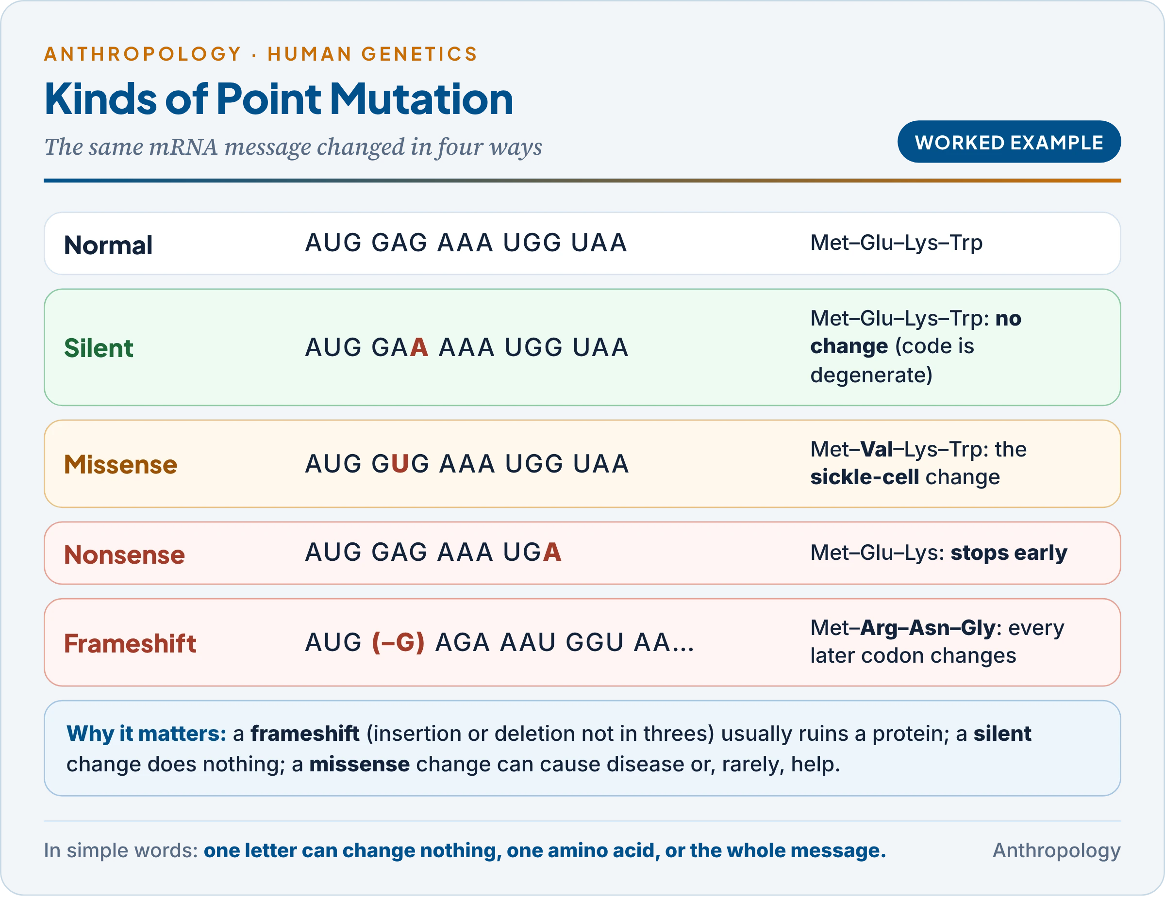
| Effect on protein | Silent, missense, nonsense, frameshift | Same amino acid; a new one (sickle cell); an early stop; frame shifted by insertion or deletion |
| Repeat expansion | Dynamic | Three-base repeat grows: CAG in Huntington’s disease |
| Origin | Spontaneous / induced | Copying errors / mutagens |
- Mutagens: physical (X-rays break DNA; heat raises the rate; ultraviolet light fuses neighbouring thymines); chemical (base analogues, alkylating agents such as mustard gas, tobacco smoke); biological (some viruses, transposons).
- Repair: proofreading, mismatch repair and excision repair fix most damage; in xeroderma pigmentosum, a recessive failure of excision repair, sunlight causes skin cancers.
6.3 Single-gene mutation disorders in man
The table lists the best-known single-gene disorders; the last column shows why several are public-health issues in India.
| Disorder | Gene (chromosome) | Inheritance | Mutation and note |
|---|---|---|---|
| Sickle-cell anaemia | HBB (11) | Autosomal recessive | Glu → Val; tribal belt of India; national mission |
| Beta-thalassaemia | HBB (11) | Autosomal recessive | Reduced beta-globin; carrier screening in several Indian communities |
| Cystic fibrosis | CFTR (7) | Autosomal recessive | Commonest: loss of one amino acid (three bases); frequent in Europeans |
| Tay-Sachs disease | HEXA (15) | Autosomal recessive | Lysosomal enzyme lost; historically common in Ashkenazi Jews |
| Phenylketonuria | PAH (12) | Autosomal recessive | Enzyme missing; newborn screening and diet prevent brain damage |
| Huntington’s disease | HTT (4) | Autosomal dominant | CAG repeat expansion; late onset |
| Achondroplasia | FGFR3 (4) | Autosomal dominant | Mostly new mutations, often from older fathers |
| Haemophilia A; G6PD deficiency | F8, G6PD (X) | X-linked recessive | Affect mainly males; G6PD deficiency frequent in some Indian groups |
6.4 Implications of mutation in evolution
- The ultimate source of new alleles: recombination reshuffles variation; only mutation creates new versions of genes.
- Neutral and adaptive change: most changes are neutral (Motoo Kimura, Unit 1.4b) and accumulate steadily, the basis of the molecular clock; a few are adaptive, such as lactase persistence and the Denisovan-derived EPAS1 variant for altitude (Unit 1.6c).
- Balanced polymorphism: a gene harmful in double dose is kept when carriers gain (sickle cell and malaria).
- Genetic load: hidden recessive mutations surface more often in consanguineous marriages (Unit 9.3).
- A tracer of history: shared mutations in mitochondrial DNA and the Y chromosome define haplogroups that map human migrations (§9).
7. Chromosomes
In simple words
Chromosomes are tightly packed bundles of DNA and protein in the nucleus. Humans have 46 in each body cell, in 23 pairs, one set from each parent. They become visible when a cell divides, and can be photographed and arranged as a karyotype.
7.1 Discovery, number and structure
- History: the German anatomist Wilhelm Waldeyer coined “chromosome” (“coloured body”) in 1888; in 1956 Joe Hin Tjio and Albert Levan counted the human number as 46, not the 48 long assumed.
- Human karyotype: 22 pairs of autosomes and one pair of sex chromosomes: 46,XX in females, 46,XY in males. Gametes are haploid (23, n); body cells diploid (46, 2n).
- Other species: chimpanzee and gorilla have 48. Human chromosome 2 formed by the fusion of two ape chromosomes: its bands match two chimpanzee chromosomes and it keeps a second, inactive centromere and telomere remnants in its middle, direct evidence of common descent.
- Packing: DNA wraps about 147 base pairs around eight histones to form a nucleosome (Roger Kornberg, 1974); further coiling compacts it about 10,000-fold at metaphase.
- Parts: two sister chromatids joined at the centromere (primary constriction), where kinetochores attach the spindle; a short arm (p) and a long arm (q); telomeres at the ends; satellites on acrocentric chromosomes carrying the ribosomal RNA genes that form the nucleolus.
- Euchromatin and heterochromatin: loosely packed, active euchromatin against densely packed, mostly inactive heterochromatin, either permanent (around centromeres, most of the Y) or facultative (the inactive X of the Barr body).
7.2 Types by centromere position
The table lists the four shapes with human examples; humans have no telocentric chromosomes.
| Type | Centromere | Human examples |
|---|---|---|
| Metacentric | Middle; equal arms | 1, 3, 16, 19, 20 |
| Submetacentric | Off-centre | 2, 4–12, 17, 18, X |
| Acrocentric | Near one end; tiny p arm with satellites | 13, 14, 15, 21, 22, Y |
| Telocentric | At the end | None in humans |
Two acrocentric chromosomes can fuse at their centromeres (a Robertsonian translocation), which causes some inherited cases of Down syndrome.
7.3 Karyotype
- Method: white blood cells are grown, stopped at metaphase with colchicine (which blocks the spindle), swollen, spread, stained and photographed; chromosomes are arranged in pairs by size and centromere (the Denver groups A–G, 1960).
- Banding: G-banding (Giemsa stain) and Q-banding (quinacrine; Torbjörn Caspersson, 1970) give each chromosome a barcode of bands, so missing, extra or moved segments can be seen.
- Uses: diagnosis of numerical and structural aberrations, prenatal testing and species comparison (Unit 9.4).
8. Cell Division
In simple words
Cells multiply by dividing. In mitosis one body cell becomes two identical cells, which is how we grow and heal. In meiosis, which happens only when eggs and sperm are made, one cell divides twice to make four cells, each with half the chromosomes and a new mix of the parents’ genes.
8.1 The cell cycle
- Phases: interphase, with G1 (growth), S (DNA replication, giving two chromatids per chromosome) and G2 (preparation), then the M phase (division). Cells that stop dividing, such as most nerve cells, rest in G0. Interphase is sometimes called a “resting stage”, but the cell is busy.
- Timing: a cultured human cell divides about every 24 hours, mitosis taking about one hour.
- Control: checkpoints run by cyclins and their kinases (Leland Hartwell, Tim Hunt and Paul Nurse, Nobel 2001) stop the cycle if DNA is damaged; p53, “guardian of the genome”, halts damaged cells. Cancer is division out of control: overactive oncogenes (stuck accelerators) and lost tumour-suppressor genes (failed brakes).
8.2 Mitosis
Mitosis (named by Walther Flemming, 1882) is the equational division of body cells: one cell gives two cells with the same 46 chromosomes.
- Prophase: chromatin condenses into chromosomes of two chromatids; the nucleolus fades; the spindle forms.
- Prometaphase: the nuclear envelope breaks down; spindle fibres attach to kinetochores.
- Metaphase: chromosomes line up on the equator, the best stage for counting them.
- Anaphase: centromeres split; sister chromatids move to opposite poles.
- Telophase and cytokinesis: nuclei re-form, ending karyokinesis (nuclear division); the cytoplasm divides by a cleavage furrow (animals) or a cell plate (plants).
Significance: mitosis keeps the chromosome number and genetic content identical, driving growth and repair.
8.3 Meiosis
Meiosis (named by J.B. Farmer and J.E.S. Moore, 1905) is the reduction division that makes gametes: one diploid cell gives four haploid cells through two divisions after one round of DNA replication.
Meiosis I (reductional: homologues separate)
- Prophase I has five stages:
- Leptotene: chromosomes condense into long threads.
- Zygotene: homologous chromosomes pair (synapsis), held by the synaptonemal complex, forming bivalents.
- Pachytene: each bivalent shows four chromatids (a tetrad); crossing over, the exchange of segments between non-sister chromatids, takes place.
- Diplotene: the synaptonemal complex dissolves; homologues stay joined at chiasmata, the X-shaped points of exchange. Human oocytes wait here (dictyotene) from before birth until ovulation, sometimes for 40 years or more.
- Diakinesis: chromosomes condense fully; chiasmata move to the ends (terminalisation).
- Metaphase I: bivalents line up on the equator, each pair oriented at random.
- Anaphase I: homologues go to opposite poles; sister chromatids stay together.
- Telophase I: two haploid cells; a brief interkinesis follows without DNA replication.
Meiosis II (equational, like mitosis): in each cell centromeres split and sister chromatids separate, giving four haploid cells.
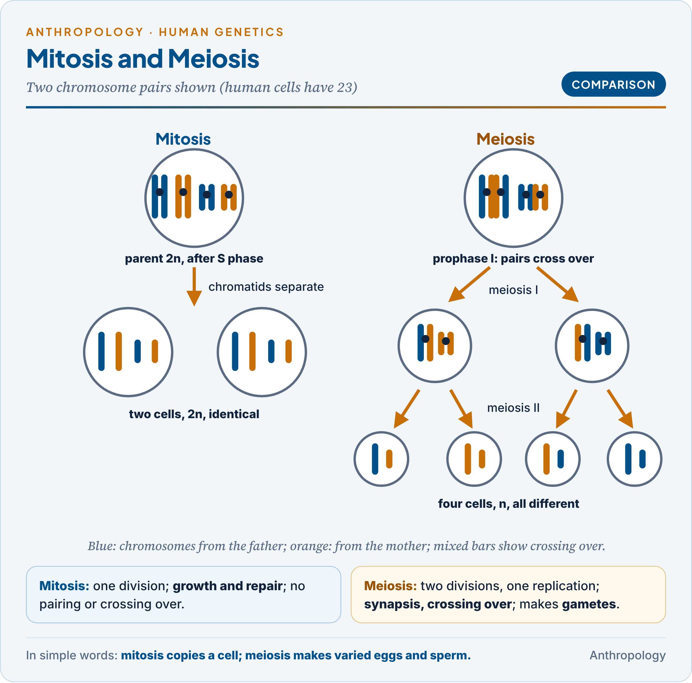
The comparison answers the commonest question on cell division; the decisive rows are pairing, crossing over and what separates at anaphase.
| Feature | Mitosis | Meiosis |
|---|---|---|
| Where | Body cells | Germ cells in testis and ovary |
| Divisions | One | Two |
| Products | Two diploid, identical cells | Four haploid, genetically different cells |
| Pairing and crossing over | No | Yes (zygotene, pachytene) |
| Anaphase separates | Sister chromatids | Homologues (I), then chromatids (II) |
| Purpose | Growth and repair | Gametes; constant number; variation |
8.4 The genetic significance of meiosis
- The physical basis of Mendel’s laws (Sutton, 1902–03): segregation is the separation of homologues at anaphase I; independent assortment is the random orientation of bivalents at metaphase I.
- Variation: random orientation alone gives 2²³, about 8.4 million, kinds of gamete per person, and two parents about 70 trillion possible children, before crossing over multiplies the number further (Worked Example 6). This is the raw material of natural selection, and the reason why no two people except identical twins are genetically alike, the basis of DNA fingerprinting.
- Errors: non-disjunction gives gametes with an extra or a missing chromosome: Down syndrome (trisomy 21), Turner (45,X) and Klinefelter (47,XXY) syndromes (Unit 9.4). Most trisomy 21 arises in maternal meiosis I, and its risk rises with maternal age, probably because oocytes wait decades in prophase I.
- Gametogenesis: in spermatogenesis a primary spermatocyte gives four sperm; in oogenesis a primary oocyte gives one ovum and polar bodies; halving here and restoring at fertilisation keeps the number at 46.
9. Nuclear and Mitochondrial DNA
In simple words
Most of our DNA is in the nucleus and comes half from each parent, reshuffled in every generation. A small extra ring of DNA inside the mitochondria comes only from the mother, unchanged except for rare mutations. That makes mitochondrial DNA a simple record of the mother’s line, which is why it was used to trace human origins to Africa.
The two genomes differ in almost every respect, and each difference has a use.
| Feature | Nuclear DNA | Mitochondrial DNA (mtDNA) |
|---|---|---|
| Size | About 3.1 billion base pairs per set | 16,569 base pairs |
| Shape | Linear, in 46 chromosomes with histones | Circular, without histones |
| Genes | About 20,000 protein-coding | 37 (13 proteins, 22 tRNAs, 2 rRNAs) |
| Copies per cell | Two of each chromosome | Hundreds to thousands |
| Inheritance | Both parents | Mother only |
| Recombination | Yes | None |
| Mutation rate | Lower | About 10 times higher, mainly in the control region |
- Why mtDNA suits evolutionary studies: it passes unchanged down the female line, so differences between people arise only from mutations that build up with time, giving one datable family tree.
- Human evolution: the “mitochondrial Eve” study (Rebecca Cann, Mark Stoneking and Allan Wilson, 1987) found the deepest lineages in Africa and traced all living mtDNA to one woman about 200,000 years ago; haplogroups M and N mark the out-of-Africa dispersal, and the Andamanese carry unique, deep M lineages (Kumarasamy Thangaraj and colleagues, 2005); Neanderthal (1997) and Denisovan (2010) mtDNA first showed these were separate lineages (Unit 1.6c).
- Uses and limits: many copies survive in old remains, which helps forensics and ancient DNA; but mtDNA records only the maternal line, so the Y chromosome and whole genomes are needed for the full picture.
- Medicine: mtDNA mutations cause diseases such as Leigh syndrome, passed from a mother to all her children. In mitochondrial donation the nucleus of an affected mother’s egg or embryo is moved into a donor egg with healthy mitochondria; the UK reported eight healthy babies born this way (Newcastle, July 2025).
Common confusion
Is a “three-parent baby” really the child of three parents? Only in a narrow sense. The baby’s nuclear DNA, with about 20,000 genes that shape its traits, comes from its mother and father as usual. The donor supplies only mitochondria, with just 37 genes for energy production. The child is genetically its parents’ child, with healthy cell “batteries” from a third person.
10. Worked Examples
In simple words
These examples show the rules of this unit at work with real numbers: base pairing, replication, the genetic code, mutation, cell division and a lethal gene. Each can be redrawn in an answer in a few lines.
1. Chargaff’s rules. A DNA sample has 30 per cent adenine. Since A = T, T = 30 per cent; G + C = 100 − 60 = 40, and G = C, so G = C = 20 per cent. Check: purines (A + G = 50) equal pyrimidines (T + C = 50). More G + C means more triple bonds and more heat-stable DNA.
2. Meselson and Stahl. Heavy (¹⁵N) bacteria are moved to light (¹⁴N) medium.
- Generation 1: 2 molecules, both hybrid (100 per cent).
- Generation 2: 4 molecules, 2 hybrid and 2 light (50 per cent).
- Generation 3: 8 molecules, 2 hybrid and 6 light (25 per cent).
- Rule: the two original strands are never lost, so the hybrid fraction after n generations is 2 / 2ⁿ. A conservative model would instead give separate heavy and light bands after generation 1.
3. Why a triplet code? With 4 bases, words of length k number 4^k: 4 for k = 1, 16 for k = 2 (fewer than 20 amino acids) and 64 for k = 3, of which 61 code for amino acids and 3 are stops, so the code is degenerate.
4. From gene to protein. Template DNA (3′ to 5′): TAC CTC TTT ATT → mRNA (5′ to 3′): AUG GAG AAA UAA → Met–Glu–Lys, then stop. The coding strand reads ATG GAG AAA TAA, the mRNA with T for U.
5. One sequence, four mutations. Start with mRNA AUG GAG AAA UGG UAA (Met–Glu–Lys–Trp–stop).
| Change | New mRNA | Protein | Type |
|---|---|---|---|
| GAG → GAA | AUG GAA AAA UGG UAA | Met–Glu–Lys–Trp | Silent |
| GAG → GUG | AUG GUG AAA UGG UAA | Met–Val–Lys–Trp | Missense (the sickle-cell change) |
| UGG → UGA | AUG GAG AAA UGA | Met–Glu–Lys, then stop | Nonsense |
| First G of GAG deleted | AUG AGA AAU GGU AA… | Met–Arg–Asn–Gly… | Frameshift |
The table shows why frameshifts are usually the most damaging and silent changes harmless: the reading frame matters more than any single letter.
6. Chromosomes and DNA through the cycle (n = 23; C = DNA of one haploid set).
| Stage | Chromosomes | Chromatids each | DNA |
|---|---|---|---|
| G1 body cell | 46 | 1 | 2*C* |
| G2 (after S phase) | 46 | 2 | 4*C* |
| Each daughter of mitosis | 46 | 1 | 2*C* |
| Each cell after meiosis I | 23 | 2 | 2*C* |
| Each gamete after meiosis II | 23 | 1 | 1*C* |
Independent assortment gives 2²³ = 8,388,608 kinds of gamete per parent, and 2²³ × 2²³, about 7 × 10¹³ (70 trillion), possible children of one couple, before crossing over.
7. A lethal gene. Yellow × yellow mice (Yy × Yy; Cuénot, 1905) should give 1 YY : 2 Yy : 1 yy. YY embryos die, so the living offspring are 2 yellow : 1 non-yellow, in litters about a quarter smaller. The gene is dominant for colour but recessive for lethality; a departure from 3 : 1 reveals it.
Key Terms
| Term | Meaning in simple words |
|---|---|
| Eukaryote | An organism whose cells have a true nucleus |
| Nucleotide | The unit of DNA or RNA: phosphate, sugar and base |
| Complementary | Matching by base pairing: A with T (or U), G with C |
| Semi-conservative replication | Copying in which each new DNA keeps one old strand |
| Okazaki fragments | Short pieces in which the lagging strand is made |
| Transcription / translation | Copying DNA into RNA / reading RNA to build a protein |
| Codon / anticodon | Three mRNA bases for one amino acid / the matching bases on tRNA |
| Intron / exon | Part of a gene cut out of the RNA / part kept |
| Operon | A group of bacterial genes switched on and off together |
| Epigenetic | A change in gene activity without a change in DNA sequence |
| Lethal gene | A gene that kills before reproductive age |
| Missense / nonsense / frameshift | A new amino acid / an early stop / a shifted reading frame |
| Karyotype | A cell’s chromosomes photographed and arranged in pairs |
| Chiasma | The X-shaped point where homologues exchanged segments |
| Non-disjunction | Failure of chromosomes to separate in division |
Contemporary Developments
- A complete personal genome (August 2026): scientists assembled both parental copies of every chromosome of a reference human genome from telomere to telomere, adding over 900 million DNA letters missing from earlier benchmarks, at about 5,000 dollars (Adam Phillippy and colleagues).
- Base editing in human embryos (June–July 2026): two studies used base editing, which changes a single DNA letter, to edit donated IVF embryos precisely, reopening the debate on germline editing.
- One-shot gene editing for cholesterol (September 2026): a single infusion of a CRISPR therapy switching off the ANGPTL3 gene halved “bad” cholesterol for a year in a first trial of 15 patients (Cleveland Clinic; New England Journal of Medicine).
- India’s sickle-cell drive (July 2026): the National Sickle Cell Anaemia Elimination Mission (launched July 2023) had made over 7.24 crore screenings (Ministry of Tribal Affairs); BIRSA 101, India’s first indigenous CRISPR therapy for sickle-cell disease (CSIR-IGIB), was launched in November 2025, with first trials expected in 2027.
Diagrams to Master
- Topic map: cell → DNA → replication → protein synthesis → gene → mutation → chromosomes → division.
- Double helix: backbone, A=T (2 bonds), G≡C (3 bonds), 5′ and 3′ ends, 2 nm, 3.4 nm per turn.
- Replication fork: helicase, primase, leading strand, Okazaki fragments, ligase.
- Protein synthesis: transcription → splicing → mRNA → ribosome (A, P, E) → protein.
- Mutation types: one sequence with silent, missense, nonsense, frameshift.
- Mitosis and meiosis: 2*n* → two 2*n*; 2*n* → bivalents with chiasmata → four n.
Revision Grid
| Scholar | Work (year) | Key idea |
|---|---|---|
| Schleiden, Schwann; Virchow | 1838–39; 1855 | Cell theory |
| Garrod | 1902–08 | Inborn errors of metabolism |
| Avery, MacLeod and McCarty | 1944 | DNA is the genetic material |
| Watson and Crick | Nature (1953) | Double helix |
| Meselson and Stahl | 1958 | Semi-conservative replication |
| Crick | 1958 | Central dogma |
| Jacob and Monod | 1961 | Lac operon |
| Nirenberg, Khorana and Holley | 1961–66 | Genetic code |
| Neel | 1962 | Thrifty genotype |
| Tjio and Levan | 1956 | 46 human chromosomes |
| Sharp and Roberts | 1977 | Split genes |
| Cann, Stoneking and Wilson | 1987 | Mitochondrial Eve |
Previous Year Questions
Unit 1.7: The biological basis of life
- 2026Write notes on: Distinguishing features of nuclear and mitochondrial DNA.
- 2025Write notes on: Mendelian and non-Mendelian traits.
- 2025Write notes on: Mitochondrial DNA and human evolution.
- 2024Write notes on: Lethal and sublethal genes.
- 2024Write notes on: Single-gene mutation disorders in man.
- 2023Write notes on: Gene expression.
- 2019Write a note in 150 words on Implications of mutation in evolution.
- 2009Write a note on Thrifty genotype.
- 2008Write a note on Protein Synthesis.
- 1992Write a note on Genes.
- 1988Give an outline of the anthropological perspective of genetics and -critically evaluate the genetic significance of meiotic cell division.
Related questions from other units and Paper II
- 2026Unit 9.4: Numerical chromosomal aberrations can be due to abnormalities in autosomes as well as sex chromosomes. Elaborate in terms of the effects these have on human body, giving suitable examples.
- 2026Unit 6.2: Write an account on the prevalence of sickle cell disease among the tribal populations of India. What are the initiatives undertaken by the affected states to tackle with this disease?
- 2024Unit 9.4: What is meant by karyotype? How does its analysis help in diagnosis of the chromosomal aberrations in man?
- 2023Unit 9.4: Describe the causes of structural abnormalities of chromosomes with suitable examples.
- 2018Unit 9.4: Write a note in 150 words on Categorize Genes that influence Human Survival.
- 2012Unit 9.2: What are lethal and sub-lethal genes ?Explain.
- 2010Unit 9.4: Write a note in 150 words on Oncogenes.
- 1998Unit 9.3: Write a note on Mutation.
- 1987Unit 9.2: Discuss how the laws of heredity propounded by Mendel can be understood in the context of meiotic cell division and their application on Man.